

MULTIMODAL IMAGING OF PHOTIC MACULOPATHY FROM ARC WELDING
- PMID: 30260904
- PMCID: PMC6435440
- DOI: 10.1097/ICB.0000000000000823
MULTIMODAL IMAGING OF PHOTIC MACULOPATHY FROM ARC WELDING
Abstract
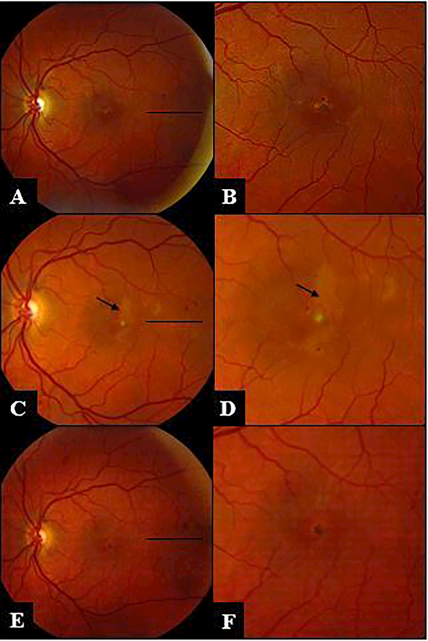
Purpose: Photic retinal toxicity induced by exposure to arc welding can lead to irreversible vision loss. Serial multimodal imaging is characterized in a patient with outer retinal damage secondary to welder's maculopathy.
Methods: A single case was retrospectively reviewed.
Results: Spectral domain optical coherence tomography acutely revealed disruption of the ellipsoid zone, hyperreflective bands through the outer nuclear layer, and outer retinal cavitation consistent with phototoxicity. Subsequently, disruption and hypertrophy of the subfoveal retinal pigment epithelium developed. Autofluorescence depicted central hypoautofluorescence.
Conclusion: We report serial multimodal imaging in welder's maculopathy to better characterize the evolution of lesions. Multimodal imaging including spectral domain optical coherence tomography in arc welding phototoxicity may share features with other forms of phototoxicity such as hand-held laser maculopathy.
Conflict of interest statement
The authors do not have any conflict of interest.
Figures


References
-
- Terrien F Des trouble visual provoque por l’electricete, Arch. Ophthalomol. 1902; 22:692–696.
-
- Bureau of Labor Statistics, U.S. Department of Labor. Welders, Cutters, Solderers, and Brazers. Occupational Outlook Handbook, 2016–17 Edition. December 17, 2015. https://www.bls.gov/ooh/production/welders-cutters-solderers-and-brazers... (visited January 31, 2017).
-
- Stokkermans TJ, Dunbar MT (1998) Solar retinopathy in a hospital-based primary care clinic. J Am Optom Assoc 1998;69(10):625–636 - PubMed
-
- Vukicevic M, Heriot W. Phototoxic maculopathy associated with arc welding: clinical findings and associated functional vision impairment. Clin Experiment Ophthalmol. 2008. October;36(7):695–7. - PubMed
-
- Mainster MA, Turner PL: Retinal injuries from light: Mechanisms, Hazards and Prevention. Edited by: Ryan SJ, Hinton DR, Schachat AP, Wilkinson P. In Retina. Volume 2. 4th edition. St Louis. Mosby Elsevier Publishers; 2006;1857–1870.
Publication types
MeSH terms
Grants and funding
LinkOut - more resources
Full Text Sources
Other Literature Sources
Medical
