. 2018 Sep 27;13(9):e0204450.
doi: 10.1371/journal.pone.0204450. eCollection 2018.

Characterization of a leukocidin identified in Staphylococcus pseudintermedius

Affiliations

Characterization of a leukocidin identified in Staphylococcus pseudintermedius

Mohamed A Abouelkhair et al. PLoS One. .

Abstract

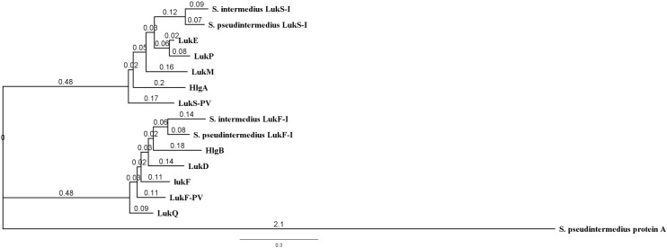
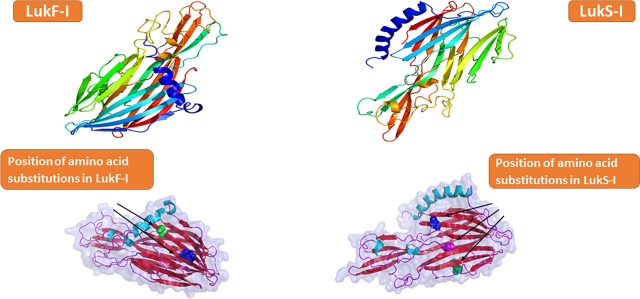
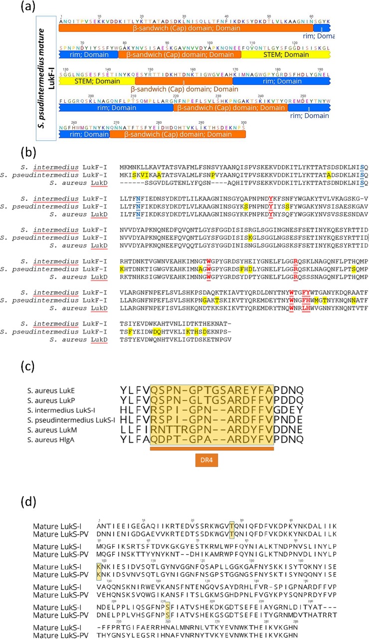
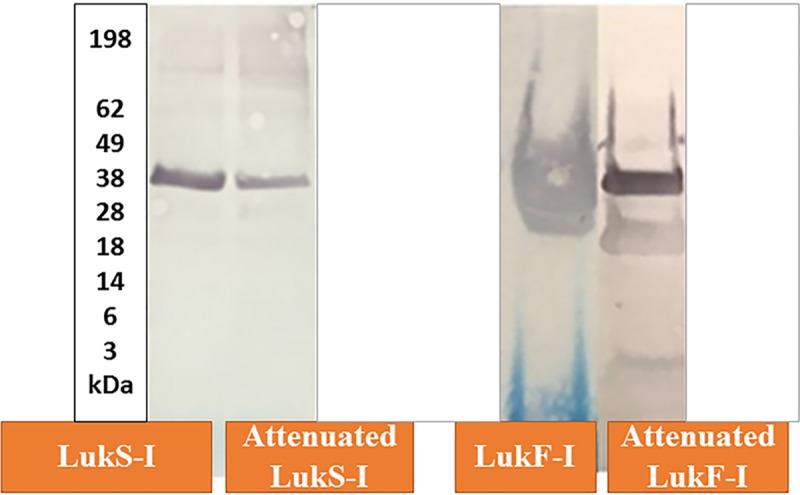
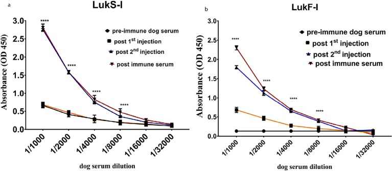
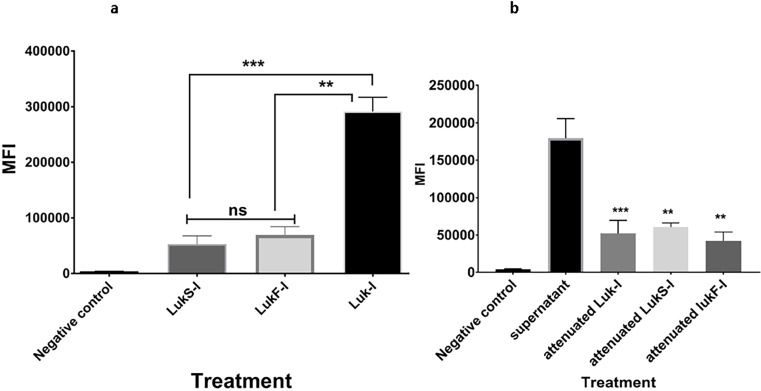
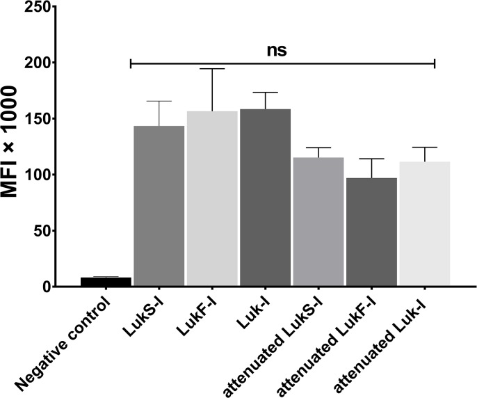
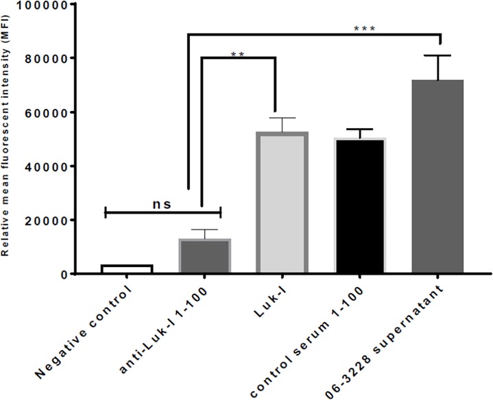
Bacterial infections from Staphylococcus pseudintermedius are the most common cause of skin infections (pyoderma) affecting dogs. Two component pore-forming leukocidins are a family of potent toxins secreted by staphylococci and consist of S (slow) and F (fast) components. They impair the innate immune system, the first line of defense against these pathogens. Seven different leukocidins have been characterized in Staphylococcus aureus, some of which are host and cell specific. Through genome sequencing and analysis of the S. pseudintermedius secretome using liquid chromatography mass spectrometry we identified two proteins, named "LukS-I" and "LukF-I", encoded on a degenerate prophage contained in the genome of S. pseudintermedius isolates. Phylogenetic analysis of LukS-I components in comparison to the rest of the leukocidin family showed that LukS-I was most closely related to S. intermedius LukS-I, S. aureus LukE and LukP, whereas LukF-I was most similar to S. intermedius LukF-I S. aureus gamma hemolysin subunit B. The killing effect of recombinant S. pseudintermedius LukS-I and LukF-I on canine polymorphonuclear leukocytes was determined using a flow cytometry cell permeability assay. The cytotoxic effect occurred only when the two recombinant proteins were combined. Engineered mutant versions of the two-component pore-forming leukocidins, produced through amino acids substitutions at selected points, were not cytotoxic. Anti-Luk-I produced in dogs against attenuated proteins reduced the cytotoxic effect of native canine leukotoxin which highlights the importance of Luk-I as a promising component in a vaccine against canine S. pseudintermedius infections.


Conflict of interest statement

We have the following interests. We have a patent application for PCT/US18/46281 "Staphylococcus pseudintermedius virulence factor compositions." This non-provisional application was filed by the University of Tennessee and is not associated with any transfer or sale of rights or ownership or any revenue. There are no further patents, products in development or marketed products to declare. This does not alter our adherence to all the PLOS ONE policies on sharing data and materials, as detailed online in the guide for authors.

Figures

Fig 1
Fig 1. Phylogenetic tree based on amino acid sequences of mature leucocidins produced by S. aureus, S. intermedius and S. pseudintermedius.
S. pseudintermedius LukS-I is closely related to S. intermedius LukS-I, S. aureus LukE and LukP. S. pseudintermedius LukF-I is closely related to S. intermedius LukF-I and S. aureus gamma hemolysin subunit B. S. pseudintermedius protein A was used as an outgroup.
Fig 2
Fig 2. Ribbon representation of Wild-type and attenuated S. pseudintermedius LukS-I and LukF-I proteins.
a, Color-ramped from the N terminus (blue) to the C terminus (red). The output structure was generated with Pymol.
Fig 3
Fig 3. Unique residues in S and F-components of S. pseudintermedius Luk-I may shape the protein function and specificity.
A, domain structure of S. pseudintermedius LukF-I consist of β-sandwich (cap) domain highlighted in orange color (1–60, 79–107, 147–169, 220–246 and 569–300), stem highlighted in yellow (108–146) and rim highlighted in blue (61–78, 170–219 and 247–268). B, Multiple sequence alignment (MSA) of S. intermedius and S. pseudintermedius LukF-I and S. aureus LukD. Geneious version 11.0.3 was used to generate the alignment. Residues unique to S. pseudintermedius LukF-I are highlighted yellow, residues identified as important for phospholipid interaction are shown in red and underlined text and residues important for oligomerization is highlighted in blue. C, MSA of amino acid sequences of the DR4 region (highlighted in yellow) in the rim domain (an important region for S-component receptor binding) of S. aureus LukE, HlgA, LukM and LukP, S. pseudintermedius LukS-I and S. intermedius LukS-I. The DR4 regions of S. aureus LukP and LukS-I of S. pseudintermedius and S.intermedius are almost identical, whilst that of LukM, HlgA and LukE are considerably different. D, MSA of amino acid sequences of S. pseudintermedius LukS-I and S. aureus LukS-PV showing the critical residues T28, K99 and S211 (highlighted in yellow) that interact with the corresponding F-component.
Fig 4
Fig 4. Western blot of recombinant S. pseudintermedius wild-type and attenuated LukS-I and LukF-I with HRP-conjugated anti-6xhis tag monoclonal antibody.
The Molecular weights of LukS-I, LukF-I, attenuated LukS-I and attenuated LukF-I determined in western blots using pre-stained Protein Standard (x 1,000) were of the expected sizes 39.43, 39.12, 37.27 and 37.59 kDa, respectively.
Fig 5
Fig 5. Canine antibody against attenuated LukS-I and LukF-I react with recombinant wild-type LukS-I and LukF-I.
Antibodies against S. pseudintermedius wild-type LukS-I and LukF-I were detected using an indirect ELISA. Recombinant S. pseudintermedius LukS-I and LukF-I proteins were coated on ELISA plates, then incubated with two-fold serially diluted serum from dog vaccinated with the same proteins. High reactivity with LukS-I and LukF-I was seen from sera collected two weeks after 3rd injetions of attenuated LukS-I (P <0.0001****) and LukF-I (P <0.0001****) compared to pre-injection sera. The values represent averages from three independent experiments.
Fig 6
Fig 6. Cytotoxic effect of S. pseudintermedius recombinant Luk-I on canine PMNs.
PMN permeability assay using Sytox Green to detect the cytotoxic effect of wild-type Luk-I on canine PMNs. (A) Luk-I significantly induced PMN killing after 30 min compared to that with wild-type LukS-I (P = 0.0330***) and LukF-I (P = 0.0468**). (B) A1:2 S. pseudintermedius 06–3228 supernatant dilution significantly induced PMN killing after 30 min compared to that with and attenuated LukS-I (P = 0.0276**), attenuated LukF-I (P = 0.0268**) and attenuated Luk-I treatments (P = 0.0044***). The mean fluorescent intensity (MFI) of all treatment were calculated based on average values from three independent experiments. (*P < 0.05 was considered significant). ns–Not significant.
Fig 7
Fig 7. S. pseudintermedius wild-type and attenuated Luk-I binding to canine PMNs.
Biotin labelled recombinant wild type and attenuated LukS-I and LukF-I were incubated with canine PMNs and their binding was detected using FITC conjugated avidin. MFI of the blank, wild-type and attenuated proteins were calculated based on average values from three independent experiments. All of the recombinant proteins bind to canine PMNs with no significant difference (ns).
Fig 8
Fig 8. Dog antibody raised against attenuated LukS-I and LukF-I protects canine PMNs from the cytotoxic effect of Luk-I.
The PMN permeability assay was performed. Luk-I preincubated with dog anti-Luk-I resulted in a significant reduction in the mean fluorescent intensity (MFI) compared with that of Luk-I treatment alone (P = 0.0036**) and with that of 06–3228 supernatant treatment alone (P = 0.0002***).

References

    1. Weese JS, van Duijkeren E. Methicillin-resistant Staphylococcus aureus and Staphylococcus pseudintermedius in veterinary medicine. Vet Microbiol. 2010;140(3–4):418–29. 10.1016/j.vetmic.2009.01.039 . - DOI - PubMed
    1. Videla R, Solyman SM, Brahmbhatt A, Sadeghi L, Bemis DA, Kania SA. Clonal Complexes and Antimicrobial Susceptibility Profiles of Staphylococcus pseudintermedius Isolates from Dogs in the United States. Microb Drug Resist. 2018;24(1):83–8. 10.1089/mdr.2016.0250 . - DOI - PubMed
    1. Solyman SM, Black CC, Duim B, Perreten V, van Duijkeren E, Wagenaar JA, et al. Multilocus sequence typing for characterization of Staphylococcus pseudintermedius. J Clin Microbiol. 2013;51(1):306–10. 10.1128/JCM.02421-12 ; PubMed Central PMCID: PMCPMC3536184. - DOI - PMC - PubMed
    1. Videla R, Solyman SM, Brahmbhatt A, Sadeghi L, Bemis DA, Kania SA. Clonal Complexes and Antimicrobial Susceptibility Profiles of Staphylococcus pseudintermedius Isolates from Dogs in the United States. Microb Drug Resist. 2017. 10.1089/mdr.2016.0250 . - DOI - PubMed
    1. Moodley A, Damborg P, Nielsen SS. Antimicrobial resistance in methicillin susceptible and methicillin resistant Staphylococcus pseudintermedius of canine origin: literature review from 1980 to 2013. Vet Microbiol. 2014;171(3–4):337–41. 10.1016/j.vetmic.2014.02.008 . - DOI - PubMed

Publication types

MeSH terms

LinkOut - more resources