. 2018 Oct:123:185-197.
doi: 10.1016/j.yjmcc.2018.09.008. Epub 2018 Sep 24.

Phospholamban regulates nuclear Ca2+ stores and inositol 1,4,5-trisphosphate mediated nuclear Ca2+ cycling in cardiomyocytes

Affiliations

Phospholamban regulates nuclear Ca2+ stores and inositol 1,4,5-trisphosphate mediated nuclear Ca2+ cycling in cardiomyocytes

Mu Chen et al. J Mol Cell Cardiol. 2018 Oct.

Abstract

Aims: Phospholamban (PLB) is the key regulator of the cardiac Ca2+ pump (SERCA2a)-mediated sarcoplasmic reticulum Ca2+ stores. We recently reported that PLB is highly concentrated in the nuclear envelope (NE) from where it can modulate perinuclear Ca2+ handling of the cardiomyocytes (CMs). Since inositol 1,4,5-trisphosphate (IP3) receptor (IP3R) mediates nuclear Ca2+ release, we examined whether the nuclear pool of PLB regulates IP3-induced nuclear Ca2+ handling.

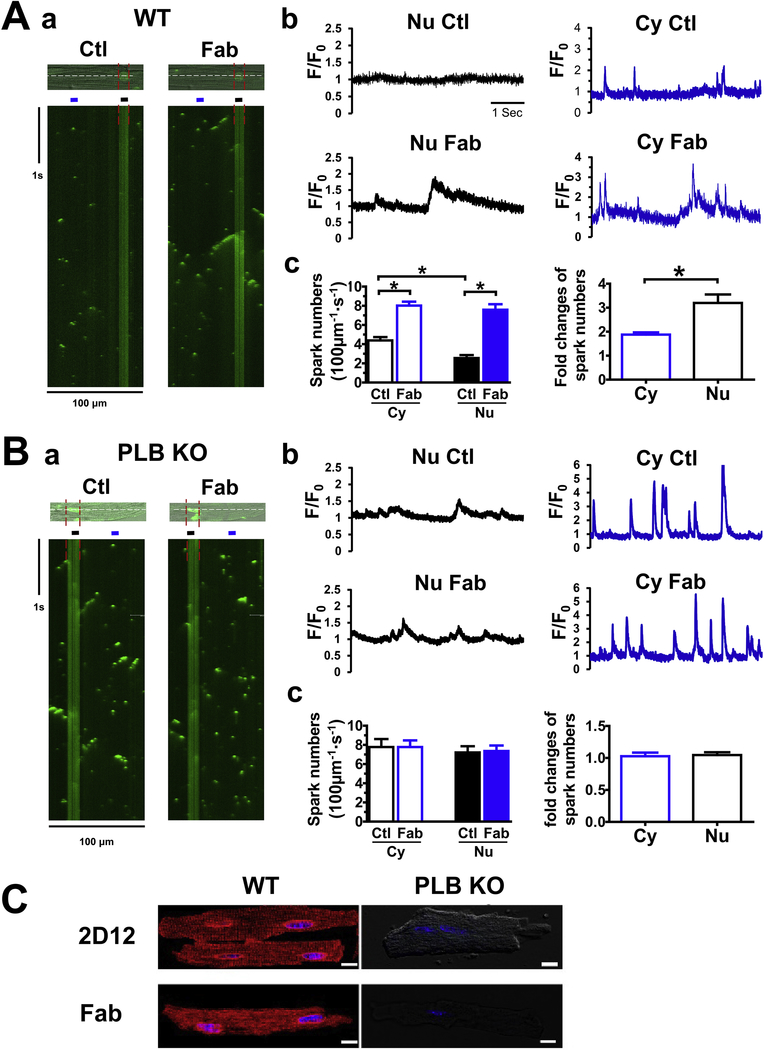
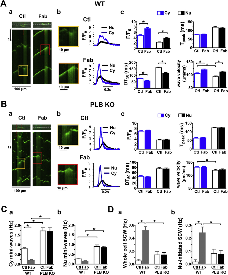
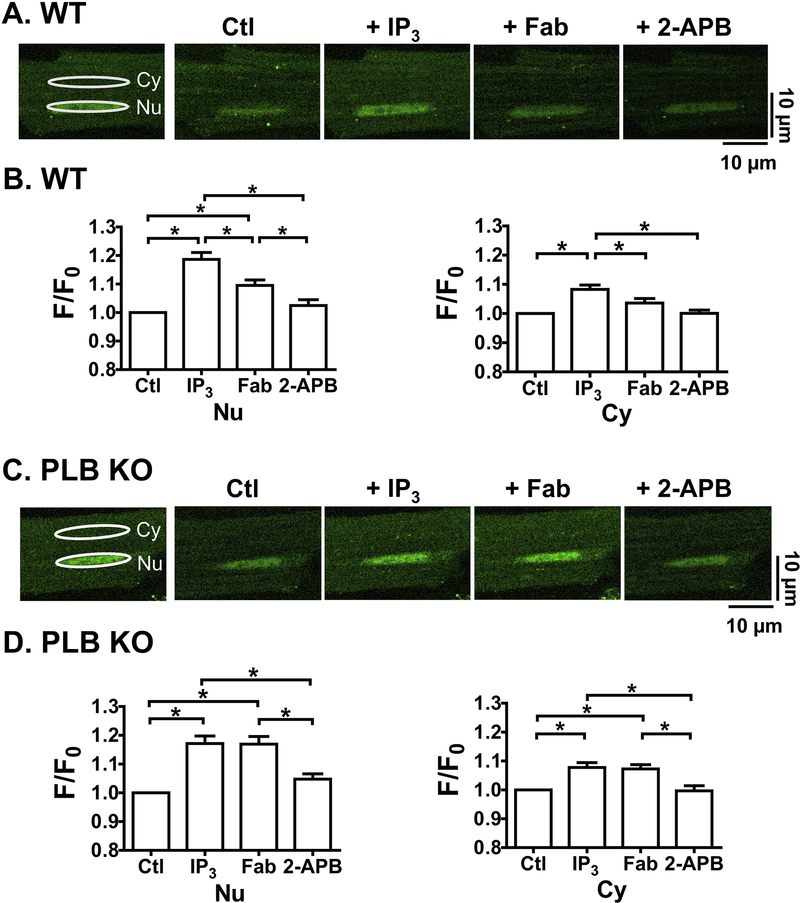
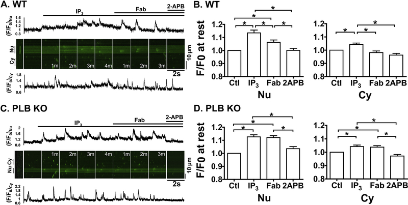
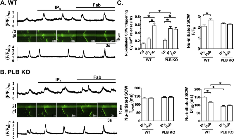
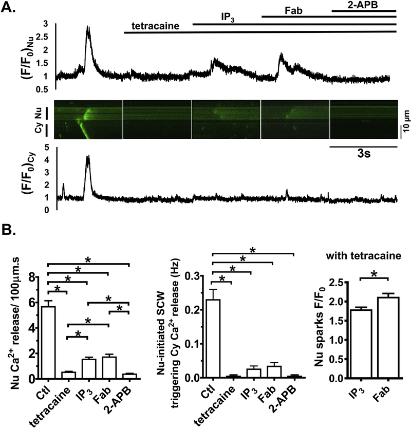
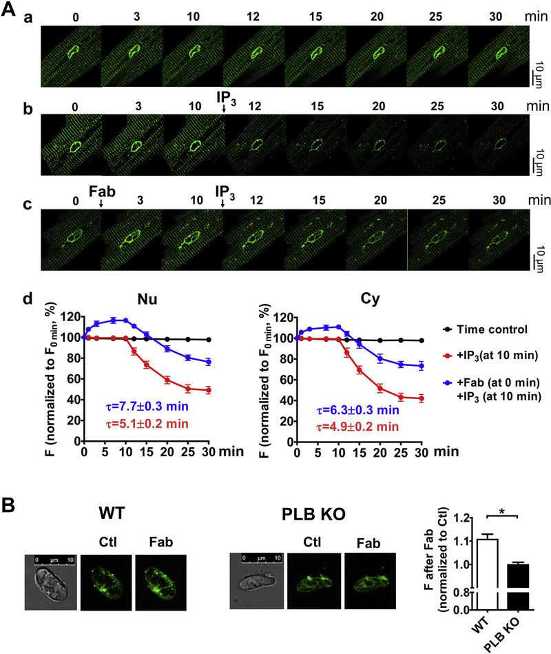
Methods and results: Fluo-4 based confocal Ca2+ imaging was performed to measure Ca2+ dynamics across both nucleus and cytosol in saponin-permeabilized CMs isolated from wild-type (WT) or PLB-knockout (PLB-KO) mice. At diastolic intracellular Ca2+ ([Ca2+]i = 100 nM), the Fab fragment of the monoclonal PLB antibody (anti-PLB Fab) facilitated the formation and increased the length of spontaneous Ca2+ waves (SCWs) originating from the nuclear region in CMs from WT but not from PLB-KO mice. We next examined nuclear Ca2+ activities at basal condition and after sequential addition of IP3, anti-PLB Fab, and the IP3R inhibitor 2-aminoethoxydiphenyl borate (2-APB) at a series of [Ca2+]i. In WT mice, at 10 nM [Ca2+]i where ryanodine receptor (RyR2) based spontaneous Ca2+ sparks rarely occurred, IP3 increased fluorescence amplitude (F/F0) of overall nuclear region to 1.19 ± 0.02. Subsequent addition of anti-PLB Fab significantly decreased F/F0 to 1.09 ± 0.02. At 50 nM [Ca2+]i, anti-PLB Fab not only decreased the overall nuclear F/F0 previously elevated by IP3, but also increased the amplitude and duration of spark-like nuclear Ca2+ release events. These nuclear Ca2+ releases were blocked by 2-APB. At 100 nM [Ca2+]i, IP3 induced short SCWs originating from nucleus. Anti-PLB Fab transformed those short waves into long SCWs with propagation from the nucleus into the cytosol. In contrast, neither nuclear nor cytosolic Ca2+ dynamics was affected by anti-PLB Fab in CMs from PLB-KO mice in all these conditions. Furthermore, in WT CMs pretreated with RyR2 blocker tetracaine, IP3 and anti-PLB Fab still increased the magnitude of nuclear Ca2+ release but failed to regenerate SCWs. Finally, anti-PLB Fab increased low Ca2+ affinity mag-fluo 4 fluorescence intensity in the lumen of NE of nuclei isolated from WT but not in PLB-KO mice.

Conclusion: PLB regulates nuclear Ca2+ handling. By increasing Ca2+ uptake into lumen of the NE and perhaps other perinuclear membranes, the acute reversal of PLB inhibition decreases global Ca2+ concentration at rest in the nucleoplasm, and increases Ca2+ release into the nucleus, through mechanisms involving IP3R and RyR2 in the vicinity.

Keywords: 1,4,5-Trisphosphate receptor; Calcium signaling; Cardiomyocyte; Nuclear Ca(2+) dynamics; Nuclear membranes; Phospholamban; Sarcoplasmic reticulum Ca(2+) cycling.


Figures

Figure 1.
Figure 1.. The effect of anti-PLB Fab on intracellular Ca2+ release in nuclear and cytoplasmic regions of CMs isolated from WT (A) or PLB-KO mice (B).
a. representative confocal line-scan Ca2+ images using Fluo-4 Ca2+ indicator were obtained in the same permeabilized mouse CM (top) before (Ctl) and after addition of 100 μg/ml anti-PLB Fab (Fab). Nucleus is between red lines. Scan-line (white) is across cytosol and nucleus. Ca2+ concentration was 50 nM. b. Traces showed intensity of fluorescent signals (F/F0) across the cytosol and nucleus (regions indicated by lines in a). c. Bar graphs showing spark frequency in the cytoplasmic and perinuclear regions, and fold of increase after addition of anti-PLB Fab. * indicates p<0.05 vs control (average of 12 CMs from 5 mice). C. Confocal immunofluorescence images showing 2D12 and anti-PLB Fab conjugated with Alexa Fluor-594 staining CMs from WT, but not from PLB-KO mice. Similar staining was obtained from at least 6 CMs isolated from WT or PLB-KO mice.
Figure 2.
Figure 2.. The effect of anti-PLB Fab on initiation of the spontaneous Ca2+ waves in cytoplasmic and perinuclear regions of CMs from WT (A) or PLB-KO mice (B).
a. representative confocal line-scan Ca2+ images using Fluo-4 Ca2+ indicator were obtained in the same permeabilized mouse CM (top) before (Ctl) and after addition of 100 μg/ml anti-PLB Fab (Fab). Nucleus is between red lines. Scan-line (white) is over cytosol and nucleus. Ca2+ concentration was 100 nM. b. Magnified region showing spontaneous Ca2+ waves (SCWs). Traces showed intensity of fluorescent signals (F/F0) of SCWs. c. Bar graphs showing characteristics of SCWs. C, D. Bar graphs showing frequency of mini-waves and long SCWs initiated at cytoplasmic and perinuclear regions. * indicates p<0.05 vs control (average of 12 CMs from 5 WT or 12 CMs from 5 PLB KO mice, respectively).
Figure 3.
Figure 3.. The effect of ET-1 (100 nM) on Ca2+ transients in cytoplasmic and perinuclear regions of CMs isolated from WT (A) or PLB-KO mice (B).
a. representative traces of Ca2+ transients in cytoplasmic and perinuclear (between red lines) regions of CMs. b and c, intensity profiles and biophysical parameters of Ca2+ transients in cytoplasmic and perinuclear regions of CMs. Each Ca transient have its own diastolic Ca level diastolic Ca in the absence of ET was used for F0 determination. * indicates p<0.05 vs control (average of 10 CMs from 5 WT or 10 CMs from 5 PLB KO mice, respectively).
Figure 4.
Figure 4.. Anti-PLB Fab affects IP3-induced nuclear Ca2+ releases at rest in WT (A, B) but not in PLB-KO (C, D).
Representative 2D confocal images show fluo-4 signals in permeabilized CMs. [Ca2+]I = 10 nM. A.C, Scan Images for nuclear (Nu) and cytosolic (Cy) regions show fluo-4 signals and intensity (F/F0) at control condition (Ctl) and 3 min after sequential addition of IP3 (10 μM), anti-PLB Fab (100μg/ml) and IP3R blocker 2-APB (10 μM). White ellipses show the identical regions of interest for detecting fluorescence intensity in Nu and Cy. B.D, plots show F/F0 at rest for Nu (left panels) and Cy (right panels) in each condition. * indicates p<0.05. n=12 CMs, 6 mice for WT; n= 12 CMs, 6 mice for PLB-KO.
Figure 5.
Figure 5.. Anti-PLB Fab affects IP3-induced nuclear Ca2+ releases at 50nM [Ca2+]i in WT (A,B) but not in PLB-KO (C,D).
Representative line-scan confocal images show fluo-4 signals in permeabilized CMs. [Ca2+]i,=50 nM. A, C. Scan Images (2 sec) and traces for nuclear (Nu, upper panels) and cytosolic (Cy, lower panels) regions show fluo-4 signals and intensity profiles (F/F0) at basal condition (Ctl) and after sequential addition of IP3 (10 μM), anti-PLB Fab (100μg/ml), and 2-APB (10 μM). M indicates minutes after addition of the reagents. B.D, Bar graphs show F/F0 at rest for Nu (left panels) and Cy (right panels) in each condition. * indicates p<0.05. n=12 CMs, 6 mice for WT; n= 13 CMs, 6 mice for PLB-KO.
Figure 6.
Figure 6.. Anti-PLB Fab affects IP3-induced SCWs originated from nuclear regions in WT (A) but not in PLB-KO (B).
Representative line-scan confocal images show fluo-4 signals in permeabilized CMs.

[Ca2+]i,=100nM. A, B. Scan images (3 sec) and traces for cytosolic (Cy, lower panels) and nuclear (Nu, upper panels) regions show fluo-4 signals and intensity profiles (F/F0) at basal (Ctl) and after sequential addition of IP3 (10 μM) and anti-PLB Fab (100μg/ml). M indicates minutes after addition of the reagents. C. Plot shows the frequency and kinetic parameters of nuclear initiated SCWs with triggering cytosolic Ca2+ release. * indicates p<0.05. n=15 CMs, 7 mice for WT; n= 14 CMs, 7 mice for PLB-KO.
Fig. 7.
Fig. 7.. Effects of anti-PLB Fab with pretreatment of tetracaine and IP3 in WT.
A. Representative line-scan confocal images and intensity profiles (F/F0) of fluo-4 signals in cytoplasmic and nuclear regions at baseline and 3 minutes after sequential addition of tetracaine (0.5 mM), IP3 (10 μM), anti-PLB Fab (100 μg/ml), and 2-APB (10 μM). B. Bar graphs show characteristics of Ca2+ release. n= 12 CMs from 6 WT mice.
Fig. 8.
Fig. 8.. Effects of anti-PLB Fab on Ca2+ concentration in the lumen of NE and SR.
Permeabilized CMs or isolated cardiac nuclei from both WT and PLB-KO mice were loaded with mag-fluo-4 and imaged. A. Representative 2D confocal images of mag-fluo-4 signals in the NE and SR from permeabilized dog CMs at a. baseline; b. after addition of IP3 (10 μM); and c. sequential addition of anti-PLB Fab (100 μg/ml), and IP3 (10 μM). d. graphs show time-dependent intensity profiles (F/F0min) after treatments. n= 6 CMs from 2 dogs. B. Representative 2D confocal images of mag-fluo-4 signals in the NE in isolated cardiac nuclei from WT and PLB-KO mice at control (Ctl) and after addition of anti-PLB Fab (Fab). Bar graph shows the mag-fluo-4 intensity ratios after addition of anti-PLB Fab.

References

    1. Simmerman HK, Jones LR, Phospholamban: protein structure, mechanism of action, and role in cardiac function, Physiol. Rev 78(4) (1998) 921–47. - PubMed
    1. MacLennan DH, Kranias EG, Phospholamban: a crucial regulator of cardiac contractility, Nat. Rev. Mol. Cell. Biol 4(7) (2003) 566–77. - PubMed
    1. Kranias EG, Hajjar RJ, Modulation of cardiac contractility by the phospholamban/SERCA2a regulatome, Circ Res 110(12) (2012) 1646–60. - PMC - PubMed
    1. Kranias EG, Hajjar RJ, The Phospholamban Journey 4 Decades After Setting Out for Ithaka, Circ Res 120(5) (2017) 781–783. - PMC - PubMed
    1. Luo W, Grupp IL, Harrer J, Ponniah S, Grupp G, Duffy JJ, Doetschman T, Kranias EG, Targeted ablation of the phospholamban gene is associated with markedly enhanced myocardial contractility and loss of beta-agonist stimulation, Circ Res 75(3) (1994) 401–9. - PubMed

Publication types