Review
. 2018 Dec:185:240-275.
doi: 10.1016/j.biomaterials.2018.09.028. Epub 2018 Sep 17.

Bone physiology as inspiration for tissue regenerative therapies

Affiliations
Review

Bone physiology as inspiration for tissue regenerative therapies

Diana Lopes et al. Biomaterials. 2018 Dec.

Abstract

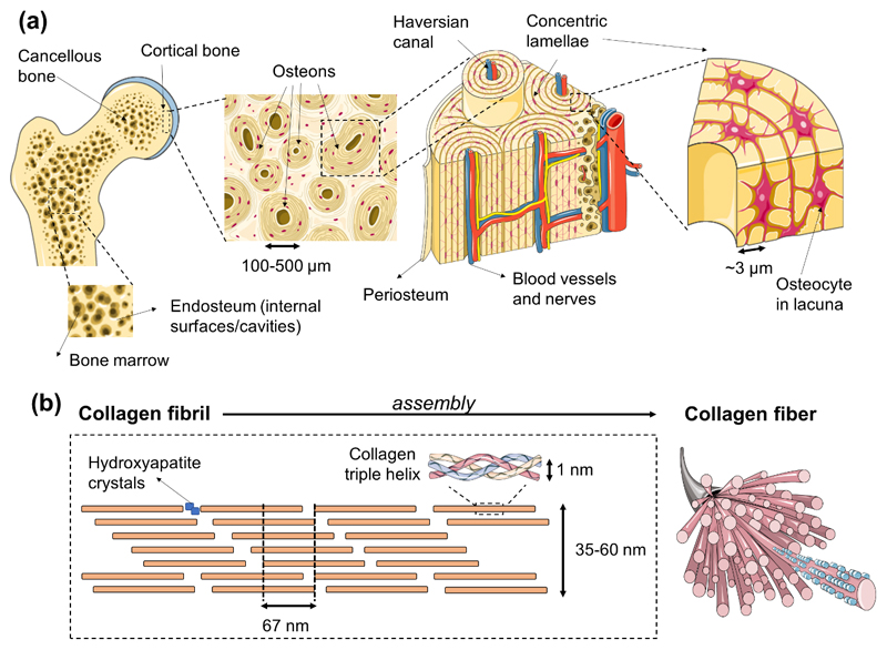
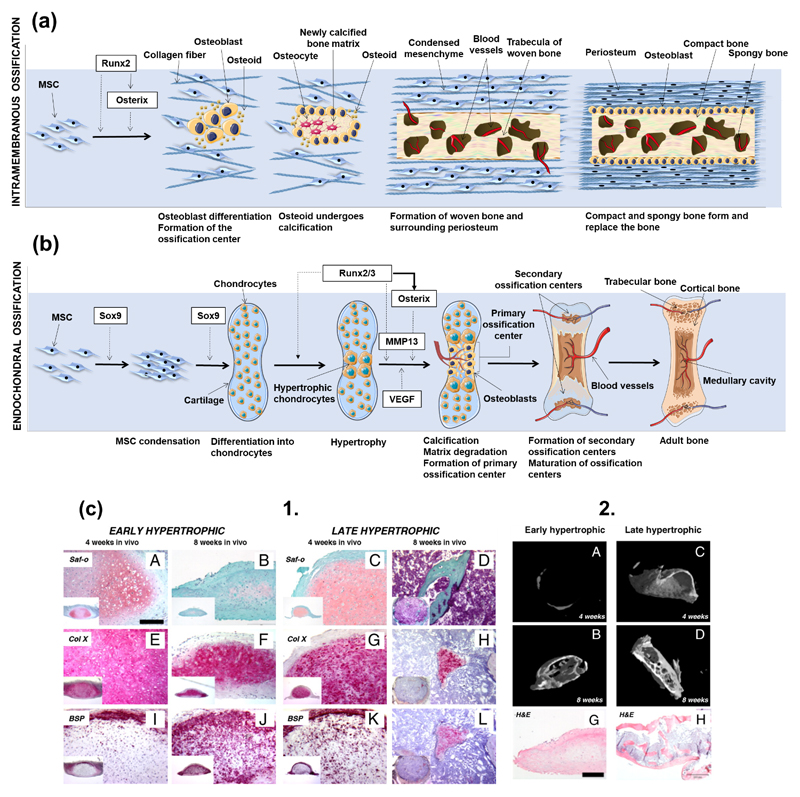
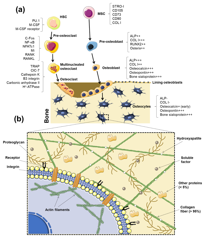
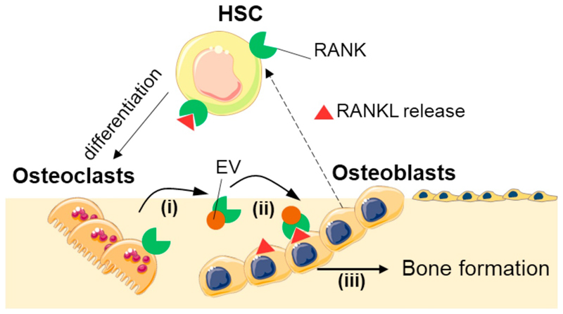
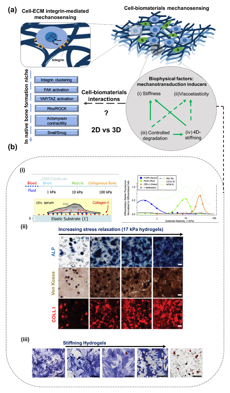
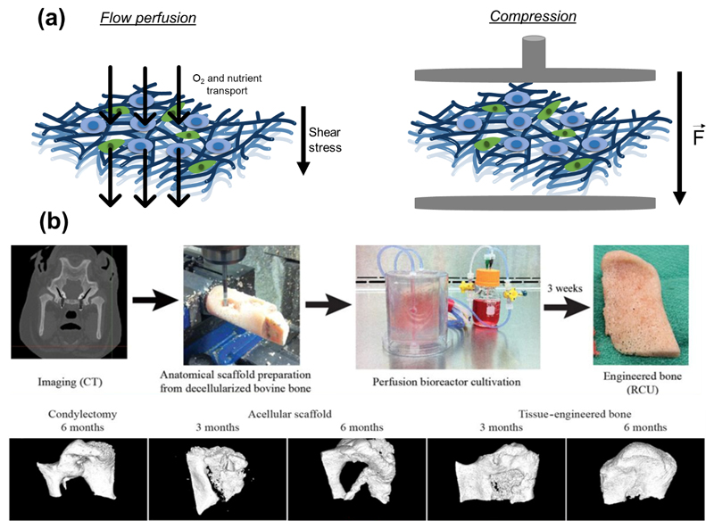
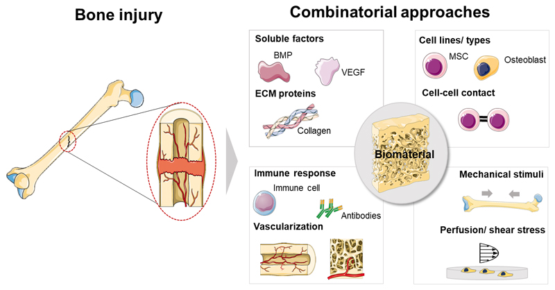
The development, maintenance of healthy bone and regeneration of injured tissue in the human body comprise a set of intricate and finely coordinated processes. However, an analysis of current bone regeneration strategies shows that only a small fraction of well-reported bone biology aspects has been used as inspiration and transposed into the development of therapeutic products. Specific topics that include inter-scale bone structural organization, developmental aspects of bone morphogenesis, bone repair mechanisms, role of specific cells and heterotypic cell contact in the bone niche (including vascularization networks and immune system cells), cell-cell direct and soluble-mediated contact, extracellular matrix composition (with particular focus on the non-soluble fraction of proteins), as well as mechanical aspects of native bone will be the main reviewed topics. In this Review we suggest a systematic parallelization of (i) fundamental well-established biology of bone, (ii) updated and recent advances on the understanding of biological phenomena occurring in native and injured tissue, and (iii) critical discussion of how those individual aspects have been translated into tissue regeneration strategies using biomaterials and other tissue engineering approaches. We aim at presenting a perspective on unexplored aspects of bone physiology and how they could be translated into innovative regeneration-driven concepts.

Keywords: Biomaterials; Biomimetics; Bone microenvironment; Bone physiology.


Figures

Figure 1
Figure 1
Interscale representation of bone. (a) A macroscopic-to-microscopic view of cancellous and cortical bone. Bone marrow lies in the cavities of cancellous bone, which are lined by the endosteum structure. Tightly packed osteons integrate cortical tissue, which is covered by the periosteum membrane. Osteons are formed by Harvasian canals, which contain blood vessels and nerve tissue, surrounded by concentric lamellae that show thicknesses of circa 3 μm. Osteocytes reside in the osteon inside lacuna structures. (b) Bone tissue is constituted at the nanometric scale by collagen fibers that comprise assembled collagen triple helix structures that give rise to the collagen fibril, with a characteristic periodic spacing of 67 nm, and gaps of 40 nm where the mineral component of bone is located.
Figure 2
Figure 2
(a) Schematic representation of intramembranous ossification. At an initial stage, MSCs cluster and differentiate into osteoblasts, forming the ossification center. Runx2 is deeply involved in the regulation of osteogenic differentiation, either directly or by inducing the late expression of Osterix. Osteoblasts start to produce the osteoid, which calcifies in few days. Osteoblasts trapped into the calcified matrix differentiate into osteocytes. Vascularized mesenchyme condenses on the external area of the woven bone, generating the periosteum. The woven bone is produced, with vascularized internal spaces that will form the marrow cavity. The surface of trabeculae is filled with matrix forming the compact bone. Spongy bone persists at the inner part. (b) Schematic representation of endochondral ossification. After condensation, MSCs starts to differentiate into chondrocytes, generating a cartilage template. Chondrocytes in the middle of the cartilage become hypertrophic. Sox9 and Runx2/3 are indispensable transcription factors for the initiation of chondrogenesis and the hypertrophy of chondrocytes, respectively. Hypertrophic chondrocytes induce vascular invasion. At this stage, Osterix functions as both a downstream and transcriptional partner of Runx2/3 during calcification and matrix degradation in cartilage, and cooperate with Runx2/3 to induce MMP13 expression. Osteoblasts differentiate from cells brought into the cartilage template with blood vessels invasion, starting to produce bone at a primary ossification center. Bone formation then spreads along the shaft forming secondary ossification centers. Finally, the adult bone, containing both trabecular and cortical bones and the medullary cavity is formed. (c) 1. Scotti et al. [74] induced endochondral bone formation in vitro using human MSCs. Hypertrophic tissue structures were implanted into nude mice to assess their ability to form trabecula bone. Both early (A-J) and later (C-L) hypertrophic samples went towards differentiation after in vivo implantation, although the latter specimen presented a more intense remodeling after 4 weeks (K), with the cartilaginous template almost resorbed after 8 weeks (L). (c) 2. Quantitative microtomography (μCT) of explants demonstrated that the deposition of mineralized matrix at the early hypertrophic samples (A-B) was reduced when compared to the late hypertrophic implanted structures (C-D). In fact, late hypertrophic samples displayed an interconnected network of trabeculae throughout the core after 8 weeks of implantation (D) [81]. Histological analysis by hematoxylin/eosin staining (G-H) revealed the presence of trabecular-like structures in the outer collar and inner core of the late, but not of the early, hypertrophic samples [81]. Figure 2(b) was adapted from Reference [81].
Figure 3
Figure 3
(a) Schematic representation of the bone cell differentiation process, generating from mesenchymal and hematopoietic stem cells. Osteoblasts descend from MSCs, which firstly differentiate into pre-osteoblasts. Osteoblasts proliferate and align in the surface of the bone while others undergo maturation into the osteocyte phenotype. HSCs differentiate into pre-osteoclasts, which become multinucleated, and finally originate mature osteoclasts responsible for bone resorption. Factors produced or expressed by different cells present in the bone niche are presented aside each cell type schematic representation. (b) A variety of factors constitute the bone extracellular environment. Biological, physical and topographical features compose a specific microenvironment capable of guiding cells into predetermined phenotypes and function. Cells interact with ECM through receptors and other proteins localized at the surface.
Figure 4
Figure 4
Representation of reciprocal interactions between osteoblasts and osteocytes. Osteoblasts (and osteocytes) release RANKL, which binds to hematopoietic stem cells, giving rise to their differentiation into osteoclasts. Ikebuchi et al. [170] proved that osteoclasts are capable of modulating osteoblasts’ ability to form new bone through the release of extracellular vesicles (EVs) that contain RANK on their surface (i). The vesicles migrate to osteoblasts’ surface (ii) leading to the binding of vesicular RANK to RANKL present at osteoblasts’ surface, and directing osteoblast to form new bone (iii). The image was adapted from an original scheme by Zaidi et al. [515].
Figure 5
Figure 5
(a) Bone mechanical microenvironment is mediated by integrin-mediated binding of bone cells to the ECM. Several pathways are described in the bone healing and homeostasis process, which include integrin clustering in the presence of stiff substrates, leading the activation of focal adhesion kinases (FAKs), which later drives the activation of the YAP/TAZ pathway. Focal adhesions also activate the RHO GTPases, which favor F-actin polymerization through the activation of RHOassociated protein kinase (ROCK) [458]. The Snail/Slug pathway is also known to occur during bone formation [464]. On biomaterials, most of these pathways (with exception to Snail/Slug) have been reported to occur in, for example, MSCs culture on 2D substrates. However, stem cells osteogenic differentiation on specific 3D setups were independent from these well-known mechanisms [487]. So far, the mechanisms driving osteogenic differentiation in 3D matrices in vitro require further exploitation towards full understanding. Interestingly, not only stiffness has been addressed as a modulator of cell response towards the osteogenic commitment. Other properties including viscoelasticity (e.g. stress relaxation) and 4D spatiotemporal degradation or stiffening have been suggested as modulators of stem cells commitment [512]. The variation of physical aspects may impact the measured properties of biomaterials overtime. In the grey circle, continuous green arrow indicates the direct impact of the variation of one factor on other overtime; discontinuous green arrows show properties that will probably influence others. (b) In 2006, the ability of 2D hydrogels’ stiffness to solely direct BMMSCs multilineage differentiation was proven for the first time [466] - example (i); stress relaxation on biomaterials with constant elastic modulus is another factor capable of directing higher production of osteogenesis-related markers by MSCs, including ALP, type I collagen and phosphate deposition (stained by von Kossa) [503] - example (ii); hydrogels with varying properties overtime showed the ability to increase the production of ALP by MSCs - example (iii) [513]. Figure 4(b) was adapted from References [466, 503, 513].
Figure 6
Figure 6
(a) Engineered devices, combining biomaterials and external stimulus allow mimicking the in vivo-occurring stimuli. Different types of bioreactors allow stimulating cells in distinct manners by mimicking the fluid shear stress through a perfusion flow method and the strain caused by compression. Figure 5(a) was produced using Servier Medical Art. (b) Perfusion flow was successfully applied for the re-cellularization of 3D scaffolds targeting facial bone reconstruction in a porcine model [514]. Indeed, such perfusion flow has proven to be effective on the homogeneous (re)population of large biomaterial and/or decellularized ECMs structures with cells of interest. The acquisition of bone defect morphology and dimensions was performed by microcomputerized tomography (μCT). Decellularized bovine bone was machined to present the exact shape of the defect, and later filled with autologous porcine ASCs, which were cultured in the 3D scaffold under perfusion flow, and later implanted in the bone defect, rendering full wound regeneration. Figure 5(b) is adapted from Reference [514].
Figure 7
Figure 7
The treatment of bone injuries may benefit from the deconstruction of the native tissue niche and on the application of concepts learnt from basic physiology to the design of efficient regenerative therapies. Although simplistic approaches based on the variation of single factors may be easier to regulate and produce with high fidelity as industrialized systems, bone’s intricate multicellular healing and homeostasis processes – characterized by fine immunological spatiotemporal coordination and unique vascular and mechanical environment – suggest that the combination of specific transversal aspects of bone physiology may hide the cue for more effective, rapid and high-quality bone formation.

References

    1. Clarke B. Normal bone anatomy and physiology. Clinical journal of the American Society of Nephrology : CJASN. 2008;3(Suppl 3):S131–9. - PMC - PubMed
    1. Quarto R, Giannoni P. Bone Tissue Engineering: Past-Present-Future. Methods in molecular biology (Clifton, NJ) 2016;1416:21–33. - PubMed
    1. Hernlund E, Svedbom A, Ivergård M, Compston J, Cooper C, Stenmark J, et al. Osteoporosis in the European Union: medical management, epidemiology and economic burden: A report prepared in collaboration with the International Osteoporosis Foundation (IOF) and the European Federation of Pharmaceutical Industry Associations (EFPIA) Archives of Osteoporosis. 2013;8:136. - PMC - PubMed
    1. Yu X, Tang X, Gohil SV, Laurencin CT. Biomaterials for Bone Regenerative Engineering. Advanced healthcare materials. 2015;4:1268–85. - PMC - PubMed
    1. Oliveira MB, Mano JF. High-throughput screening for integrative biomaterials design: exploring advances and new trends. Trends in biotechnology. 2014;32:627–36. - PubMed

Publication types

Substances

LinkOut - more resources