

Haploinsufficiency of Homeodomain Proteins Six3, Vax1, and Otx2 Causes Subfertility in Mice via Distinct Mechanisms
- PMID: 30261489
- PMCID: PMC6437011
- DOI: 10.1159/000494086
Haploinsufficiency of Homeodomain Proteins Six3, Vax1, and Otx2 Causes Subfertility in Mice via Distinct Mechanisms
Abstract
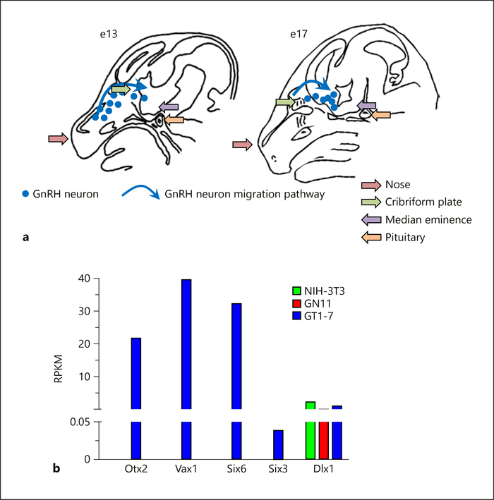
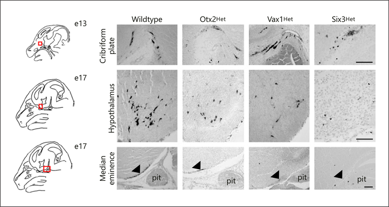
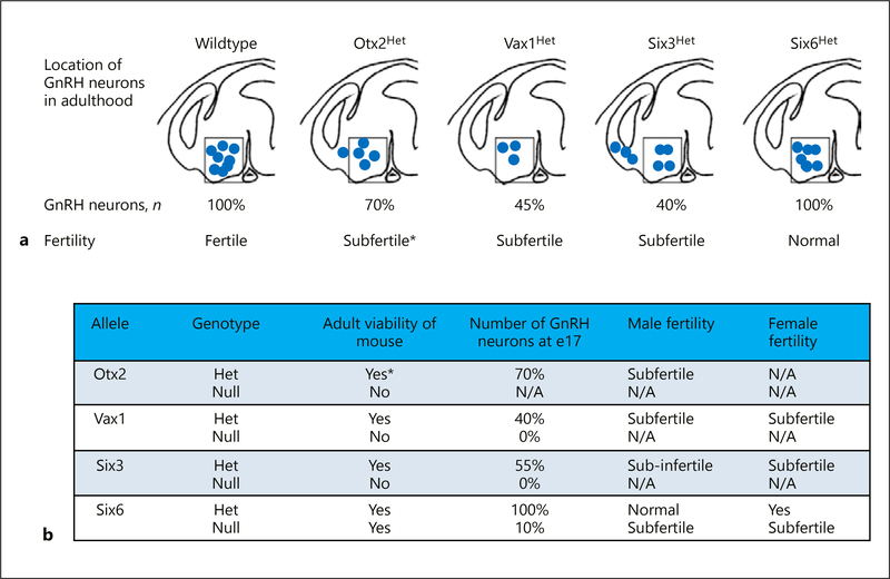
Haploinsufficiency occurs when loss of one copy of a diploid gene (hemizygosity) causes a phenotype. It is relatively rare, in that most genes can produce sufficient mRNA and protein from a single copy to prevent any loss of normal activity and function. Reproduction is a complex process relying on migration of GnRH neurons from the olfactory placode to the hypothalamus during development. We have studied 3 different homeodomain genes Otx2, Vax1, and Six3 and found that the deletion of one allele for any of these genes in mice produces subfertility or infertility in one or both sexes, despite the presence of one intact allele. All 3 heterozygous mice have reduced numbers of GnRH neurons, but the mechanisms of subfertility differ significantly. This review compares the subfertility phenotypes and their mechanisms.
Keywords: Heterozygous; Homeodomain; Infertility.
© 2018 S. Karger AG, Basel.
Conflict of interest statement
Disclosure Statement
The authors have no conflicts of interest to declare.
Figures
Similar articles
-
Haploinsufficiency of SIX3 Abolishes Male Reproductive Behavior Through Disrupted Olfactory Development, and Impairs Female Fertility Through Disrupted GnRH Neuron Migration.Mol Neurobiol. 2018 Nov;55(11):8709-8727. doi: 10.1007/s12035-018-1013-0. Epub 2018 Mar 27. Mol Neurobiol. 2018. PMID: 29589282 Free PMC article.
-
Deletion of the homeodomain gene Six3 from kisspeptin neurons causes subfertility in female mice.Mol Cell Endocrinol. 2022 Apr 15;546:111577. doi: 10.1016/j.mce.2022.111577. Epub 2022 Feb 2. Mol Cell Endocrinol. 2022. PMID: 35121076 Free PMC article.
-
Deletion of Vax1 from Gonadotropin-Releasing Hormone (GnRH) Neurons Abolishes GnRH Expression and Leads to Hypogonadism and Infertility.J Neurosci. 2016 Mar 23;36(12):3506-18. doi: 10.1523/JNEUROSCI.2723-15.2016. J Neurosci. 2016. PMID: 27013679 Free PMC article.
-
The transcription factors SIX3 and VAX1 are required for suprachiasmatic nucleus circadian output and fertility in female mice.J Neurosci Res. 2021 Oct;99(10):2625-2645. doi: 10.1002/jnr.24864. Epub 2021 Jul 2. J Neurosci Res. 2021. PMID: 34212416 Free PMC article.
-
SIX3 function in cancer: progression and comprehensive analysis.Cancer Gene Ther. 2022 Nov;29(11):1542-1549. doi: 10.1038/s41417-022-00488-9. Epub 2022 Jun 28. Cancer Gene Ther. 2022. PMID: 35764712 Review.
Cited by
-
Differential CRE Expression in Lhrh-cre and GnRH-cre Alleles and the Impact on Fertility in Otx2-Flox Mice.Neuroendocrinology. 2019;108(4):328-342. doi: 10.1159/000497791. Epub 2019 Feb 10. Neuroendocrinology. 2019. PMID: 30739114 Free PMC article.
-
The Homeodomain Transcription Factors Vax1 and Six6 Are Required for SCN Development and Function.Mol Neurobiol. 2020 Feb;57(2):1217-1232. doi: 10.1007/s12035-019-01781-9. Epub 2019 Nov 9. Mol Neurobiol. 2020. PMID: 31705443 Free PMC article.
-
Circadian Rhythms in the Neuronal Network Timing the Luteinizing Hormone Surge.Endocrinology. 2022 Feb 1;163(2):bqab268. doi: 10.1210/endocr/bqab268. Endocrinology. 2022. PMID: 34967900 Free PMC article. Review.
-
Dual Role of DLK1 in GnRH Neuron Ontogeny.Stem Cell Rev Rep. 2025 Sep 9. doi: 10.1007/s12015-025-10972-y. Online ahead of print. Stem Cell Rev Rep. 2025. PMID: 40924042
-
The homeodomain transcription factor Six3 regulates hypothalamic Pomc expression and its absence from POMC neurons induces hyperphagia and mild obesity in male mice.Mol Metab. 2024 Sep;87:101993. doi: 10.1016/j.molmet.2024.101993. Epub 2024 Jul 16. Mol Metab. 2024. PMID: 39025297 Free PMC article.
References
-
- Strauss JF, Barbieri RL. Yen and Jaffe’s Reproductive Endocrinology. 5th ed. Philadelphia (PA): Elsevier Saunders; 2004.
-
- Walters KA, Allan CM, Jimenez M, Lim PR, Davey RA, Zajac JD, et al. Female mice haploinsufficient for an inactivated androgen receptor (AR) exhibit age-dependent defects that resemble the AR null phenotype of dysfunctional late follicle development, ovulation, and fertility. Endocrinology. 2007. August; 148(8): 3674–84. - PubMed
Publication types
MeSH terms
Substances
Grants and funding
LinkOut - more resources
Full Text Sources
Other Literature Sources
Medical
