. 2018 Sep 26;10(10):392.
doi: 10.3390/toxins10100392.

Organic and Peptidyl Constituents of Snake Venoms: The Picture Is Vastly More Complex Than We Imagined

Affiliations

Organic and Peptidyl Constituents of Snake Venoms: The Picture Is Vastly More Complex Than We Imagined

Alejandro Villar-Briones et al. Toxins (Basel). .

Abstract

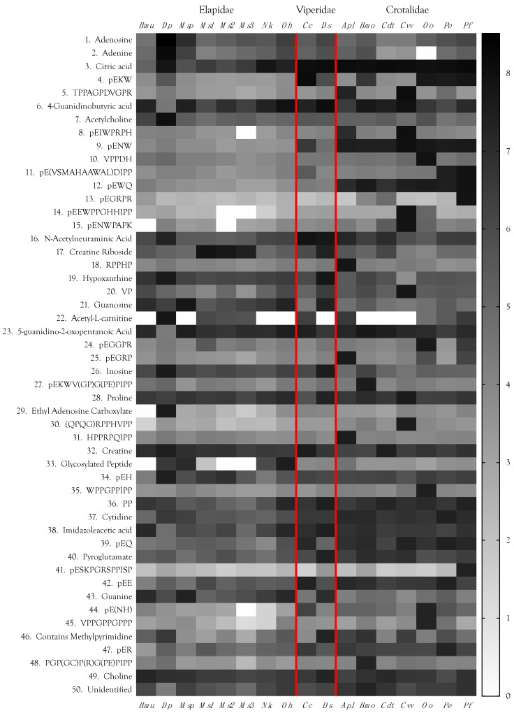
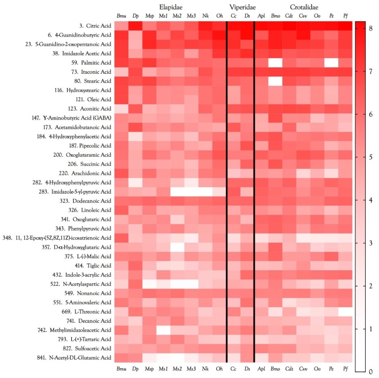
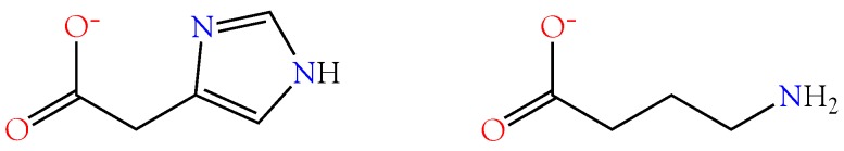
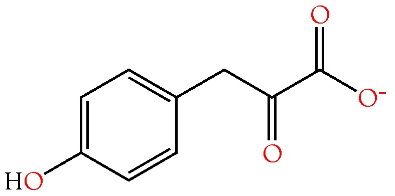
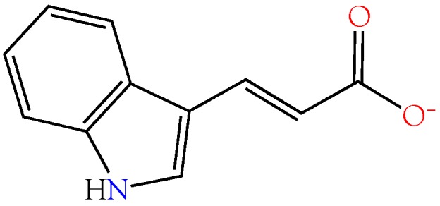
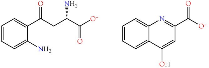
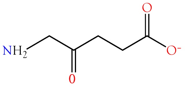
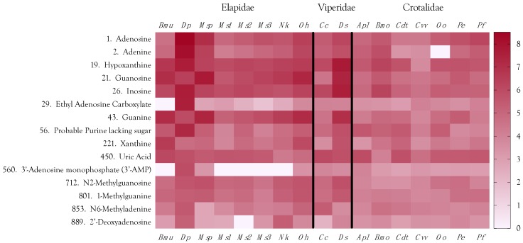
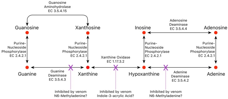
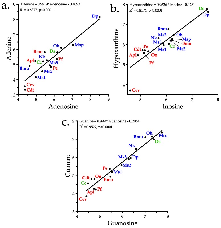
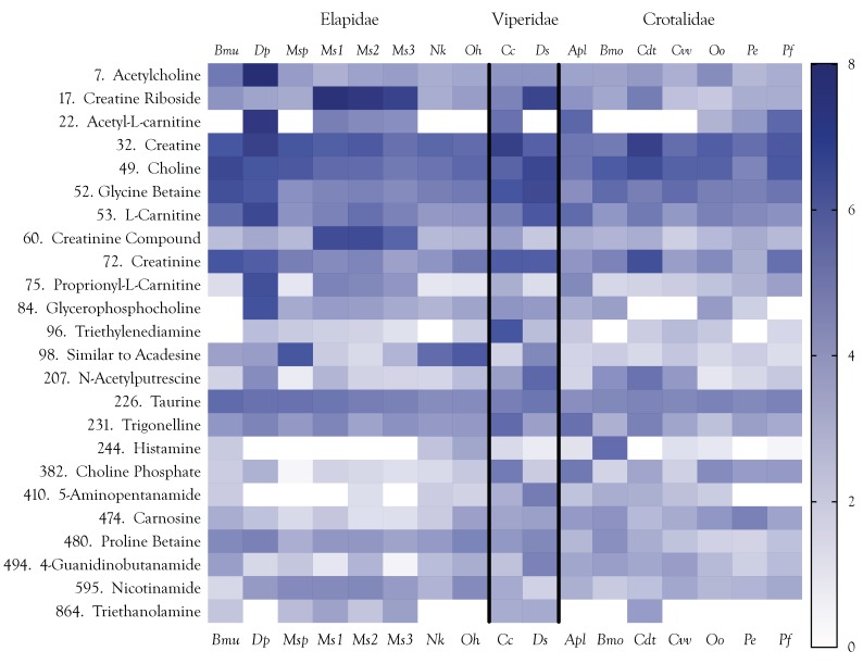
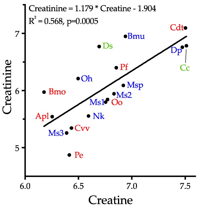
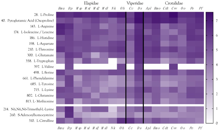
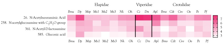
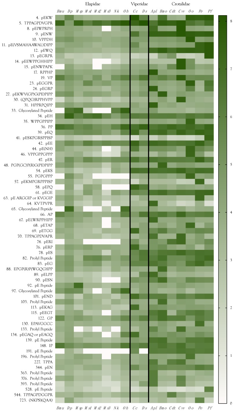
Small metabolites and peptides in 17 snake venoms (Elapidae, Viperinae, and Crotalinae), were quantified using liquid chromatography-mass spectrometry. Each venom contains >900 metabolites and peptides. Many small organic compounds are present at levels that are probably significant in prey envenomation, given that their known pharmacologies are consistent with snake envenomation strategies. Metabolites included purine nucleosides and their bases, neurotransmitters, neuromodulators, guanidino compounds, carboxylic acids, amines, mono- and disaccharides, and amino acids. Peptides of 2⁻15 amino acids are also present in significant quantities, particularly in crotaline and viperine venoms. Some constituents are specific to individual taxa, while others are broadly distributed. Some of the latter appear to support high anabolic activity in the gland, rather than having toxic functions. Overall, the most abundant organic metabolite was citric acid, owing to its predominance in viperine and crotaline venoms, where it chelates divalent cations to prevent venom degradation by venom metalloproteases and damage to glandular tissue by phospholipases. However, in terms of their concentrations in individual venoms, adenosine, adenine, were most abundant, owing to their high titers in Dendroaspis polylepis venom, although hypoxanthine, guanosine, inosine, and guanine all numbered among the 50 most abundant organic constituents. A purine not previously reported in venoms, ethyl adenosine carboxylate, was discovered in D. polylepis venom, where it probably contributes to the profound hypotension caused by this venom. Acetylcholine was present in significant quantities only in this highly excitotoxic venom, while 4-guanidinobutyric acid and 5-guanidino-2-oxopentanoic acid were present in all venoms.

Keywords: amines; amino acids; carboxylic acids; guanidinium compounds; metabolites; mono- and disaccharides; neuromodulators; neurotransmitters; peptides; purine nucleosides and bases; snake venoms.


Conflict of interest statement

The authors declare no conflict of interest.

Figures

Figure 1
Figure 1
Total ion chromatograms of negative and positive ions of metabolites and peptides from Agkistrodon piscivorus leucostoma venom. The negative ion peak that dwarfs all others is citric acid. Assuming no metabolite loss during deproteination, the metabolites and peptides separated here represent the small molecule component of ~69 µg of crude venom. Metabolites were separated on a SeQuant ZIC-pHILIC HPLC 2.1 × 150 mm column, flow rate 120 µL/min, using acetonitrile as solvent A, and 10 mM ammonium carbonate, 0.1% ammonium hydroxide in water as solvent B. Separation was done in HILIC mode, with a linear gradient from 20% to 80% solvent B in 30 min, followed by a wash for 20 min with 20% acetonitrile, 0.5 M sodium chloride in water (solvent C) and, finally, column re-equilibration with starting conditions for 15 min.
Figure 2
Figure 2
Heat map of the 50 most abundant metabolites and peptides found in 17 snake venoms, arranged in decreasing order of the maximum concentrations found among the species examined. Compound abundances represent the log10 of peak intensities of positive and negative ions combined, after subtraction of respective baselines. Logarithmic representations have the effect of compressing apparent differences, so these venoms are compositionally much more divergent than can be shown graphically. Taxonomic names: Bmu, Bungarus multicinctus; Dp, Dendroaspis polylepis; Msp, Micrurus spixii; Ms1–3, Micrurus surinamensis, 3 individuals; Nk, Naja kaouthia; Oh, Ophiophagus hannah; Cc, Cerastes cerastes; Ds, Daboia siamensis; Apl, Agkistrodon piscivorus leucostoma; Bmo, Bothrops moojeni; Cdt, Crotalus durissus terrificus; Cvv, Crotalus viridis viridis; Oo, Ovophis okinavensis; Pe, Protobothrops elegans; Pf, Protobothrops flavoviridis.
Figure 3
Figure 3
Structures of citric acid, cis-aconitic acid, and itaconic acid. cis-Aconitate is an intermediate between itaconic acid and citric acid, in the citric acid cycle. It seems probable that itaconic acid and cis-aconitic acid exist to support citric acid production in the venom glands. These tricarboxylic acids chelate divalent cations to inactivate phospholipases, metalloproteases, nucleases, and other metalloenzymes in the venom gland; however, upon injection into prey tissues, these components are immediately activated.
Figure 4
Figure 4
Organic acid abundances in snake venoms span nearly 8 orders of magnitude, based on combined positive and negative ion intensities, after subtraction of the blanks. The vast majority are unquestionably accidental venom constituents, probably resulting from cellular degradation. However, compounds with peak intensities above E06, are probably sufficiently concentrated to make substantive contributions to venom pharmacology. Baseline (noise) has been subtracted from all ion intensities. Taxonomic names: Bmu, Bungarus multicinctus; Dp, Dendroaspis polylepis; Msp, Micrurus spixii; Ms1–3, Micrurus surinamensis, 3 individuals; Nk, Naja kaouthia; Oh, Ophiophagus hannah; Cc, Cerastes cerastes; Ds, Daboia siamensis; Apl, Agkistrodon piscivorus leucostoma; Bmo, Bothrops moojeni; Cdt, Crotalus durissus terrificus; Cvv, Crotalus viridis viridis; Oo, Ovophis okinavensis; Pe, Protobothrops elegans; Pf, Protobothrops flavoviridis.
Figure 5
Figure 5
The guanidino group of L-arginine is utilized in the production of nitric oxide (NO). However, l-arginine can also be oxidized to 2-oxoarginine (2OA), a potent convulsant that exerts its effects by blocking chloride channels of GABAA and glycine receptors. With two additional enzymatic reactions, 2OA can be converted to γ-guanidinobutyric acid, which is also a convulsant. Some guanidino compounds also reduce blood pressure and suppress “fight or flight” responses in rats and aggressive behavior in cats. All of these pharmacological effects are consistent with snake envenomation strategies [4].
Figure 6
Figure 6
Imidazole-4-acetic acid, (left) an agonist of mammalian GABAA receptors. The natural agonist, γ-amino butyric acid, or GABA, is shown on the right.
Figure 7
Figure 7
Structure of 4-hydroxyphenylpyruvic acid, an inhibitor of acetylcholinesterase produced by the action of venom l-amino acid oxidase on tyrosine [93].
Figure 8
Figure 8
Structure of indole-3-acrylic acid, an inhibitor of xanthine oxidase, kynurenine aminotransferase, and d-dopachrome tautomerase.
Figure 9
Figure 9
Structures of l-kynurenine (left) and kynurenic acid (right). l-kynurenines such as quinolinic acid are excitatory, but kynurenic acid, produced from l-kynurenine by the action of kynurenine aminotransferase (KCAT1), is an inhibitor of NMDA iGluRs and α7 nAChRs. I3AA inhibits KCAT1, blocking production of both molecules by this pathway.
Figure 10
Figure 10
Structure of 5-aminolevulinic acid.
Figure 11
Figure 11
Purine nucleosides and their bases are significant constituents of elapid, viperine, and crotaline venoms. Elapid and viperine venoms contain greater quantities of them than crotaline venoms. The purine strategy of D. polylepis is particularly noteworthy. Taxonomic names: Bmu, Bungarus multicinctus; Dp, Dendroaspis polylepis; MSP, Micrurus spixii; Ms1–3, Micrurus surinamensis, 3 individuals; Nk, Naja kaouthia; Oh, Ophiophagus hannah; Cc, Cerastes cerastes; Ds, Daboia siamensis; Apl, Agkistrodon piscivorus leucostoma; Bmo, Bothrops moojeni; Cdt, Crotalus durissus terrificus; Cvv, Crotalus viridis viridis; Oo, Ovophis okinavensis; Pe, Protobothrops elegans; Pf, Protobothrops flavoviridis.
Figure 12
Figure 12
Elapid and viperine venoms contain high levels of purine nucleosides, while crotaline venoms tend to have trace quantities [4,12]. However, crotaline venoms accomplish the same objective by releasing purines from prey tissues. Venom purines isolated during this study are indicated by red dots. Interestingly, blockade of xanthine oxidase by venom indole-3-acrylic acid might drive hypoxanthine toward inosine or adenine in the venom gland; however, it is most abundant in crotaline venoms (Figure 11), which have very low purine titers. Therefore, this function seems unlikely. Perhaps it serves a similar function in prey tissues. Venoms also contain much lower levels still of N6-methyladenine, an inhibitor of both adenine and guanine deaminases; however, this compound is most abundant in mamba venom, which employs a purinergic envenomation strategy. Thus, it may support adenosine synthesis in some fashion, perhaps by blocking the backward conversion of adenine to hypoxanthine.
Figure 13
Figure 13
Concentrations of purine bases in venoms are highly correlated with concentrations of their respective nucleosides, suggesting that their primary function is to support production of the nucleosides, the roles in envenomation of which have been well characterized. Ovophis okinavensis was excluded from plot 13a because no adenine was detected in that venom. Elapids, red; viperines, green; and crotalines, blue. Taxonomic abbreviations: Apl, Agkistrodon piscivorus leucostoma; Bmo, Bothrops moojeni; Bmu, Bungarus multicinctus; Cc, Cerastes cerastes; Cdt, Crotalus durissus terrificus; Cvv, Crotalus viridis viridis; Ds, Daboia siamensis; Dp, Dendroaspis polylepis; Mss, Micrurus spixii spixii; Ms1–3, Micrurus surinamensis 1–3; Nk, Naja kaouthia; Oh, Ophiophagus hannah; Oo, Ovophis okinavensis; Pe, Protobothrops elegans; Pf, Protobothrops flavoviridis.
Figure 14
Figure 14
Various amines and cholines (quaternary amines) were identified in the 17 snake venoms. Most were present at low levels, suggesting that functional roles in envenomation are improbable. Others showed modest to high concentrations in specific taxa, but were essentially absent in others. Examples of this pattern include acetyl-l-carnitine, proprionyl-l-carnitine, triethylenediamine, histamine, and 5-aminopentanamide. Taxonomic names: Bmu, Bungarus multicinctus; Dp, Dendroaspis polylepis; Msp, Micrurus spixii; Ms1–3, Micrurus surinamensis, 3 individuals; Nk, Naja kaouthia; Oh, Ophiophagus hannah; Cc, Cerastes cerastes; Ds, Daboia siamensis; Apl, Agkistrodon piscivorus leucostoma; Bmo, Bothrops moojeni; Cdt, Crotalus durissus terrificus; Cvv, Crotalus viridis viridis; Oo, Ovophis okinavensis; Pe, Protobothrops elegans; Pf, Protobothrops flavoviridis.
Figure 15
Figure 15
Creatinine levels are well correlated with creatine levels, reflecting their metabolic link; however, they show no obvious relationship to either phylogeny or ecology, suggesting the lack of a functional role in debilitation of prey. It seems reasonable that the elevated levels of these compounds simply reflect the high rate of ATP anabolism and catabolism in the gland, resulting from the demands of protein synthesis. Elapids, red; viperines, green; and crotalines, blue. Taxonomic abbreviations: Apl, Agkistrodon piscivorus leucostoma; Bmo, Bothrops moojeni; Bmu, Bungarus multicinctus; Cc, Cerastes cerastes; Cdt, Crotalus durissus terrificus; Cvv, Crotalus viridis viridis; Ds, Daboia siamensis; Dp, Dendroaspis polylepis; Mss, Micrurus spixii spixii; Ms1–3, Micrurus surinamensis 1–3; Nk, Naja kaouthia; Oh, Ophiophagus hannah; Oo, Ovophis okinavensis; Pe, Protobothrops elegans; Pf, Protobothrops flavoviridis.
Figure 16
Figure 16
Free amino acids found in venoms, listed in the order of abundance. l-arginine serves as the precursor for nitric oxide; therefore, it potentially promotes hypotension. Proline is a major constituent in hypotensive peptides, hence its abundance in Crotalus v. viridis venom. Pyroglutamic acid (oxoproline) likewise blocks the N-terminus of crotaline and viperine hypotensive peptides; hence its greater abundance in those venoms. Trimethyl-lysine is a precursor for carnitine synthesis. Both trimethyl-lysine and proprionyl-l-carnitine are most concentrated in D. polylepis venom. High concentrations of some other amino acids are more difficult to explain. Abundance is scaled on the basis of the log10 of the total ion concentration. Taxonomic names: Bmu, Bungarus multicinctus; Dp, Dendroaspis polylepis; Msp, Micrurus spixii; Ms1–3, Micrurus surinamensis, 3 individuals; Nk, Naja kaouthia; Oh, Ophiophagus hannah; Cc, Cerastes cerastes; Ds, Daboia siamensis; Apl, Agkistrodon piscivorus leucostoma; Bmo, Bothrops moojeni; Cdt, Crotalus durissus terrificus; Cvv, Crotalus viridis viridis; Oo, Ovophis okinavensis; Pe, Protobothrops elegans; Pf, Protobothrops flavoviridis.
Figure 17
Figure 17
Relatively few mono- and disaccharides were found in these venoms and all were in the N-acetylated form. N-Acetylneuraminic acid is a common terminating sugar in the branched, asparagine-linked glycan moieties of snake venom glycoproteins. Mannose was not recorded, but it could not have been detected under the conditions were used, except at very high concentrations. Taxonomic names: Bmu, Bungarus multicinctus; Dp, Dendroaspis polylepis; Msp, Micrurus spixii; Ms1–3, Micrurus surinamensis, 3 individuals; Nk, Naja kaouthia; Oh, Ophiophagus hannah; Cc, Cerastes cerastes; Ds, Daboia siamensis; Apl, Agkistrodon piscivorus leucostoma; Bmo, Bothrops moojeni; Cdt, Crotalus durissus terrificus; Cvv, Crotalus viridis viridis; Oo, Ovophis okinavensis; Pe, Protobothrops elegans; Pf, Protobothrops flavoviridis.
Figure 18
Figure 18
All venoms examined contained peptides. Those sequenced ranged from 172–1716 Da (2–15 amino acids). Many of these were pyroglutamyl and/or prolyl peptides. Their pharmacologies are largely unknown at this point. Taxonomic names: Bmu, Bungarus multicinctus; Dp, Dendroaspis polylepis; Msp, Micrurus spixii; Ms1–3, Micrurus surinamensis, 3 individuals; Nk, Naja kaouthia; Oh, Ophiophagus hannah; Cc, Cerastes cerastes; Ds, Daboia siamensis; Apl, Agkistrodon piscivorus leucostoma; Bmo, Bothrops moojeni; Cdt, Crotalus durissus terrificus; Cvv, Crotalus viridis viridis; Oo, Ovophis okinavensis; Pe, Protobothrops elegans; Pf, Protobothrops flavoviridis.
Figure 19
Figure 19
Clustering of 17 venomous snake taxa according to identities and concentrations of the top 100 small metabolites in their venoms. (A) Dendrogram of the 17 specimens based upon similarities in their small metabolites and peptides. (B) Cluster analysis. Owing to their conspecificity and their preferred fish diet, three specimens of Micrurus surinamensis cluster tightly, well separated from everything else (Cluster 1). Cluster 2 comprises New World pitvipers, except for the viperine, Daboia siamensis, and Naja kaouthia, which is something of a dietary generalist, adults of which feed mostly on rodents and other small mammals. Micrurus spixii, which feeds upon snakes and amphisbaenians, most resembles the Ophiophagus Asian taxon, Bungarus multicinctus, in terms of venom metabolites. Ophiophagus hannah preys upon both snakes and mammals, and also falls into Cluster 3, although in the dendrogram, it clusters with N. kaouthia, C. d. terrificus, and the two viperines. The reason for this apparent discrepancy is that in the cluster analysis, only the first two of three dimensions can be shown. Cluster 4 includes Asian pitvipers and Cerastes cerastes, which sits on the border of Cluster 2. Cluster 5 contains Dendroaspis polylepis alone, the venom small metabolome of which is as unusual as its proteome. Colors in the dendrogram (A) reflect colors in the cluster analysis (B). The R script used to generate the dendrogram and to perform the cluster analysis is provided in Supplementary File 1.
Figure 19
Figure 19
Clustering of 17 venomous snake taxa according to identities and concentrations of the top 100 small metabolites in their venoms. (A) Dendrogram of the 17 specimens based upon similarities in their small metabolites and peptides. (B) Cluster analysis. Owing to their conspecificity and their preferred fish diet, three specimens of Micrurus surinamensis cluster tightly, well separated from everything else (Cluster 1). Cluster 2 comprises New World pitvipers, except for the viperine, Daboia siamensis, and Naja kaouthia, which is something of a dietary generalist, adults of which feed mostly on rodents and other small mammals. Micrurus spixii, which feeds upon snakes and amphisbaenians, most resembles the Ophiophagus Asian taxon, Bungarus multicinctus, in terms of venom metabolites. Ophiophagus hannah preys upon both snakes and mammals, and also falls into Cluster 3, although in the dendrogram, it clusters with N. kaouthia, C. d. terrificus, and the two viperines. The reason for this apparent discrepancy is that in the cluster analysis, only the first two of three dimensions can be shown. Cluster 4 includes Asian pitvipers and Cerastes cerastes, which sits on the border of Cluster 2. Cluster 5 contains Dendroaspis polylepis alone, the venom small metabolome of which is as unusual as its proteome. Colors in the dendrogram (A) reflect colors in the cluster analysis (B). The R script used to generate the dendrogram and to perform the cluster analysis is provided in Supplementary File 1.

Similar articles

Cited by

References

    1. Ganguly S.N., Malkana M.T. Indian snake venoms. II. Cobra venom: Its chemical composition, protein fractions and their physiological actions. Indian J. Med. Res. 1936;24:281–286.
    1. Devi A. The protein and nonprotein constituents of snake venoms. In: Bücherl W., Buckley E.E., Deulofeu V., editors. Venomous Animals and Their Venoms. Volume 1. Academic Press; New York, NY, USA: 1968. pp. 119–165.
    1. Bieber A.L. Metal and Nonprotein Constituents in Snake Venoms. In: Md C.-Y.L., editor. Snake Venoms. Handbook of Experimental Pharmacology; Springer; Berlin/Heidelberg, Germany: 1979. pp. 295–306.
    1. Aird S.D. Ophidian envenomation strategies and the role of purines. Toxicon. 2002;40:335–393. doi: 10.1016/S0041-0101(01)00232-X. - DOI - PubMed
    1. Fischer F.G., Dorfel H. Adenosine in the venom of puffadder Bitis arietans. Snake venoms. I. Hoppe Seylers Z. Physiol. Chem. 1954;296:232–238. doi: 10.1515/bchm2.1954.296.1.232. - DOI - PubMed

Publication types

LinkOut - more resources