

Targeting Hepatic Protein Carbonylation and Oxidative Stress Occurring on Diet-Induced Metabolic Diseases through the Supplementation with Fish Oils
- PMID: 30261666
- PMCID: PMC6213247
- DOI: 10.3390/md16100353
Targeting Hepatic Protein Carbonylation and Oxidative Stress Occurring on Diet-Induced Metabolic Diseases through the Supplementation with Fish Oils
Abstract
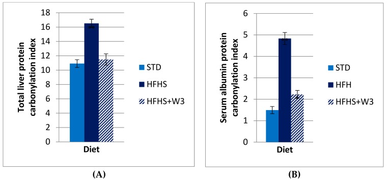
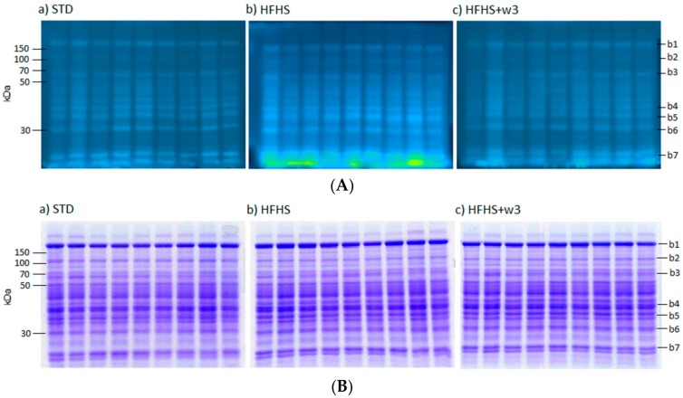
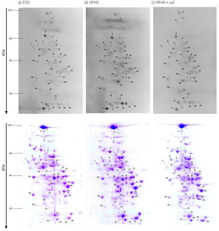
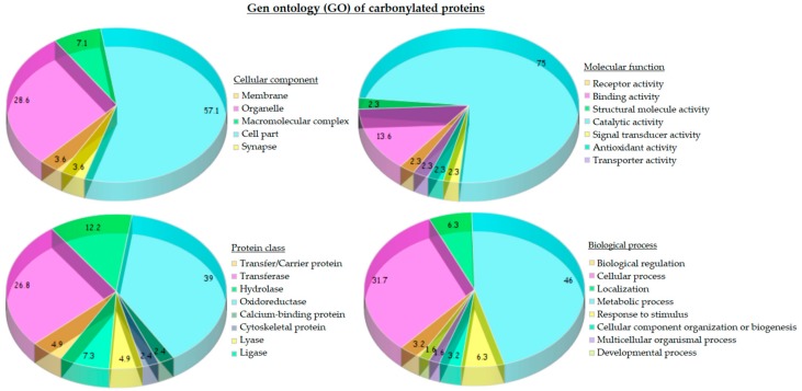
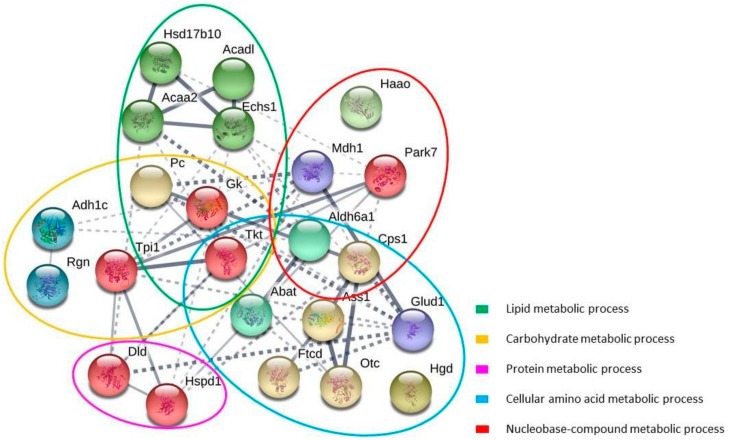
The present study addressed the ability of long-chain ω-3 polyunsaturated fatty acids (ω-3 PUFA), i.e., eicosapentaenoic acid (EPA) and docosahexaenoic acid (DHA), to ameliorate liver protein damage derived from oxidative stress and induced by consumption of high-caloric diets, typical of Westernized countries. The experimental design included an animal model of Sprague-Dawley rats fed high-fat high-sucrose (HFHS) diet supplemented with ω-3 EPA and DHA for a complete hepatic proteome analysis to map carbonylated proteins involved in specific metabolic pathways. Results showed that the intake of marine ω-3 PUFA through diet significantly decreased liver protein carbonylation caused by long-term HFHS consumption and increased antioxidant system. Fish oil modulated the carbonylation level of more than twenty liver proteins involved in critical metabolic pathways, including lipid metabolism (e.g., albumin), carbohydrate metabolism (e.g., pyruvate carboxylase), detoxification process (e.g., aldehyde dehydrogenase 2), urea cycle (e.g., carbamoyl-phosphate synthase), cytoskeleton dynamics (e.g., actin), or response to oxidative stress (e.g., catalase) among others, which might be under the control of diet marine ω-3 PUFA. In parallel, fish oil significantly changed the liver fatty acid profile given by the HFHS diet, resulting in a more anti-inflammatory phenotype. In conclusion, the present study highlights the significance of marine ω-3 PUFA intake for the health of rats fed a Westernized diet by describing several key metabolic pathways which are protected in liver.
Keywords: Sprague-Dawley rat; carbonylation; fish oils; high-fat high-sucrose diet; liver protein damage; marine omega-3 fatty acids; oxidative stress.
Conflict of interest statement
The authors declare no conflict of interest.
Figures
Similar articles
-
Modulation of the Liver Protein Carbonylome by the Combined Effect of Marine Omega-3 PUFAs and Grape Polyphenols Supplementation in Rats Fed an Obesogenic High Fat and High Sucrose Diet.Mar Drugs. 2019 Dec 30;18(1):34. doi: 10.3390/md18010034. Mar Drugs. 2019. PMID: 31906027 Free PMC article.
-
A lipidomic study on the regulation of inflammation and oxidative stress targeted by marine ω-3 PUFA and polyphenols in high-fat high-sucrose diets.J Nutr Biochem. 2017 May;43:53-67. doi: 10.1016/j.jnutbio.2017.02.007. Epub 2017 Feb 21. J Nutr Biochem. 2017. PMID: 28260647
-
Effects of long-term administration of saturated and n-3 fatty acid-rich diets on lipid utilisation and oxidative stress in rat liver and muscle tissues.Br J Nutr. 2013 Nov;110(10):1789-802. doi: 10.1017/S0007114513001311. Epub 2013 May 9. Br J Nutr. 2013. PMID: 23656726
-
Influence of Lipid Class Used for Omega-3 Fatty Acid Supplementation on Liver Fat Accumulation in MASLD.Physiol Res. 2024 Aug 31;73(Suppl 1):S295-S320. doi: 10.33549/physiolres.935396. Epub 2024 Jul 17. Physiol Res. 2024. PMID: 39016154 Free PMC article. Review.
-
Role of ω3 long-chain polyunsaturated fatty acids in reducing cardio-metabolic risk factors.Endocr Metab Immune Disord Drug Targets. 2011 Sep 1;11(3):232-46. doi: 10.2174/187153011796429817. Endocr Metab Immune Disord Drug Targets. 2011. PMID: 21831036 Review.
Cited by
-
The Effects of the Combination of Buckwheat D-Fagomine and Fish Omega-3 Fatty Acids on Oxidative Stress and Related Risk Factors in Pre-Obese Rats.Foods. 2021 Feb 4;10(2):332. doi: 10.3390/foods10020332. Foods. 2021. PMID: 33557198 Free PMC article.
-
Effects of a Fish Oil Rich in Docosahexaenoic Acid on Cardiometabolic Risk Factors and Oxidative Stress in Healthy Rats.Mar Drugs. 2021 Sep 29;19(10):555. doi: 10.3390/md19100555. Mar Drugs. 2021. PMID: 34677454 Free PMC article.
-
Dietary Marine Oils Selectively Decrease Obesogenic Diet-Derived Carbonylation in Proteins Involved in ATP Homeostasis and Glutamate Metabolism in the Rat Cerebellum.Antioxidants (Basel). 2024 Jan 15;13(1):103. doi: 10.3390/antiox13010103. Antioxidants (Basel). 2024. PMID: 38247527 Free PMC article.
-
Modulation of the Liver Protein Carbonylome by the Combined Effect of Marine Omega-3 PUFAs and Grape Polyphenols Supplementation in Rats Fed an Obesogenic High Fat and High Sucrose Diet.Mar Drugs. 2019 Dec 30;18(1):34. doi: 10.3390/md18010034. Mar Drugs. 2019. PMID: 31906027 Free PMC article.
-
Design of Fluorescent Probes for Bioorthogonal Labeling of Carbonylation in Live Cells.Sci Rep. 2020 May 6;10(1):7668. doi: 10.1038/s41598-020-64790-y. Sci Rep. 2020. PMID: 32376913 Free PMC article.
References
-
- Dichi I., Simão A.N.C., Vannucchi H., Curi R., Calder P.C. Metabolic Syndrome: Epidemiology, Pathophysiology, and Nutrition Intervention. [(accessed on 31 July 2018)]; Available online: https://www.hindawi.com/journals/jnme/2012/584541/ - PMC - PubMed
MeSH terms
Substances
LinkOut - more resources
Full Text Sources
Other Literature Sources
Medical
Research Materials
