Review
. 2018 Sep 27;19(10):2937.
doi: 10.3390/ijms19102937.

Ageing, Cellular Senescence and Neurodegenerative Disease

Affiliations
Review

Ageing, Cellular Senescence and Neurodegenerative Disease

Marios Kritsilis et al. Int J Mol Sci. .

Abstract

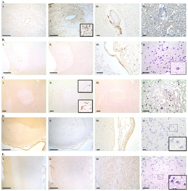
Ageing is a major risk factor for developing many neurodegenerative diseases. Cellular senescence is a homeostatic biological process that has a key role in driving ageing. There is evidence that senescent cells accumulate in the nervous system with ageing and neurodegenerative disease and may predispose a person to the appearance of a neurodegenerative condition or may aggravate its course. Research into senescence has long been hindered by its variable and cell-type specific features and the lack of a universal marker to unequivocally detect senescent cells. Recent advances in senescence markers and genetically modified animal models have boosted our knowledge on the role of cellular senescence in ageing and age-related disease. The aim now is to fully elucidate its role in neurodegeneration in order to efficiently and safely exploit cellular senescence as a therapeutic target. Here, we review evidence of cellular senescence in neurons and glial cells and we discuss its putative role in Alzheimer's disease, Parkinson's disease and multiple sclerosis and we provide, for the first time, evidence of senescence in neurons and glia in multiple sclerosis, using the novel GL13 lipofuscin stain as a marker of cellular senescence.

Keywords: Alzheimer’s disease; Parkinson’s disease; SenTraGorTM (GL13); ageing; cellular senescence; lipofuscin; multiple sclerosis; neurodegeneration; senolytics.


Conflict of interest statement

The authors declare no conflict of interest.

Figures

Figure 1
Figure 1
Lipofuscin accumulation as a marker of cellular senescence in multiple sclerosis lesions. Demyelinated lesions were identified with myelin basic protein (MBP) immunohistochemistry and were staged according to Trapp et al. (1998) [287] as acute active, chronic active or chronic inactive using human leukocyte antigen-DR isotype (HLA-DR) immunohistochemistry on serial sections from paraffin embedded postmortem tissue blocks. Lipofuscin was detected with the GL13 hybrid histochemistry-immunohistochemistry method [58]. Acute active demyelinated white matter lesion with MBP staining showing ongoing perivascular demyelination in subcortical white matter from the parietal lobe of a 73-year-old secondary progressive multiple sclerosis (SP-MS) patient (MS51) (A(i)). Infiltration with HLA-DR+ cells with macrophage morphology throughout the demyelinated parenchyma (HLA DR immunohistochemistry) (A(ii)). Perivascular infiltration with CD8+ lymphocytes (CD8 immunohistochemistry) (A(iii)). GL13 staining in acute active lesions showed lipofuscin+ cells. Although many of them were perivascularly localized, some were not, suggesting that at least some of them maybe glial cells rather than inflammatory cells (A(iv)). Chronic actively demyelinating perivenentricular white matter lesion with a fully demyelinated lesion center (lack of MBP immunoreactivity) from a 74-year-old female MS patient (MS265) (B(i)). Typically, HLA-DR immunohistochemistry of serial sections exhibited a border infiltrated by numerous macrophages whereas the lesion centre is infiltrated by ramified microglia (B(ii)). Few CD8+ lymphocytes are present perivascularly (CD8 immunohistochemistry) (B(iii)). Lipofuscin+ cells with granular staining were found in the macrophage infiltrated lesion border (B(iv)). Chronic inactive subcortical white matter demyelinated lesion (lack of MBP immunoreactivity with a well demarcated border) from the left parietal lobe of a 71-year-old female SP-MS patient (MS33) (C(i)). Ramified microglial morphology throughout the demyelinated lesion area and lesion border (HLA-DR immunohistochemistry) typical of a chronic inactive lesion (C(ii)). Decreased axonal density in the demyelinated lesion seen with 200 KDa neurofilament immunohistochemistry (C(iii)). Numerous parenchymal lipofuscin+ cells in the demyelinated white matter. Lack of HLA-DR+ macrophages from the chronic demyelinated lesion suggests that the lipofuscin+ cells are glial (C(iv)). Subpial cortical demyelination (lack of MBP immunoreactivity extending from the pial surface into the deeper cortical layers from the parietal cortex of a 71-year-old female SP-MS patient (MS33) (D(i)). HLA-DR+ ramified microglia in the demyelinated cortical lesion (D(ii)) and few CD8+ lymphocytes infiltrating the adjacent pia matter (D(iii)). Lipofuscin+ cells mostly with neuronal morphology (inset) throughout the demyelinated cortex (D(iv)). Normal appearing cortex with intact appearing cortical myelin (MBP immunohistochemistry) from the left parietal lobe of a 71-year-old female SP-MS patient (MS33) (E(i)), HLA-DR immunoreactivity revealing quiescent ramified microglia (E(ii)) and normal-appearing axonal staining with 200 kDa neurofilament immunohistochemistry on a serial section (E(iii)). GL13 staining showed numerous lipofuscin+ cells mostly with neuronal morphology (E(iv)). Scale bars represent 500 μm (A(i),A(ii),B(i),B(ii),C(i),C(ii),C(iii),D(i),D(ii),E(i),E(ii)), 50 μm (D(iii),E(iii)) or 25 μm (A(iii),A(iv), B(iii), B(iv), C(iv), D(iv), E(iv)).

Similar articles

Cited by

References

    1. Johnson I.P. Age-related neurodegenerative disease research needs aging models. Front. Aging Neurosci. 2015;7:168. doi: 10.3389/fnagi.2015.00168. - DOI - PMC - PubMed
    1. Schrijvers E.M., Verhaaren B.F., Koudstaal P.J., Hofman A., Ikram M.A., Breteler M.M. Is dementia incidence declining? Trends in dementia incidence since 1990 in the Rotterdam Study. Neurology. 2012;78:1456–1463. doi: 10.1212/WNL.0b013e3182553be6. - DOI - PubMed
    1. Rocca W.A., Petersen R.C., Knopman D.S., Hebert L.E., Evans D.A., Hall K.S., Gao S., Unverzagt F.W., Langa K.M., Larson E.B., et al. Trends in the incidence and prevalence of Alzheimer’s disease, dementia, and cognitive impairment in the United States. Alzheimers Dement. 2011;7:80–93. doi: 10.1016/j.jalz.2010.11.002. - DOI - PMC - PubMed
    1. Nussbaum R.L., Ellis C.E. Alzheimer’s Disease and Parkinson’s Disease. N. Engl. J. Med. 2003;348:1356–1364. doi: 10.1056/NEJM2003ra020003. - DOI - PubMed
    1. Lockrow J.P., Fortress A.M., Granholm A.C.E. Age-related neurodegeneration and memory loss in down syndrome. Curr. Gerontol. Geriatr. Res. 2012;2012:13. doi: 10.1155/2012/463909. - DOI - PMC - PubMed