. 2019 Jan 1;27(1):54-62.
doi: 10.4062/biomolther.2017.237.

Cis-3-O-p- hydroxycinnamoyl Ursolic Acid Induced ROS-Dependent p53-Mediated Mitochondrial Apoptosis in Oral Cancer Cells

Affiliations

Cis-3-O-p- hydroxycinnamoyl Ursolic Acid Induced ROS-Dependent p53-Mediated Mitochondrial Apoptosis in Oral Cancer Cells

Ching-Ying Wang et al. Biomol Ther (Seoul). .

Abstract

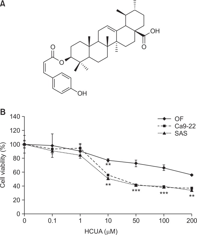
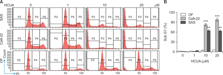
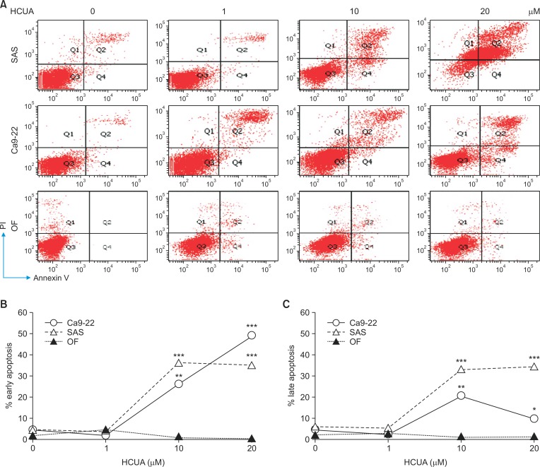
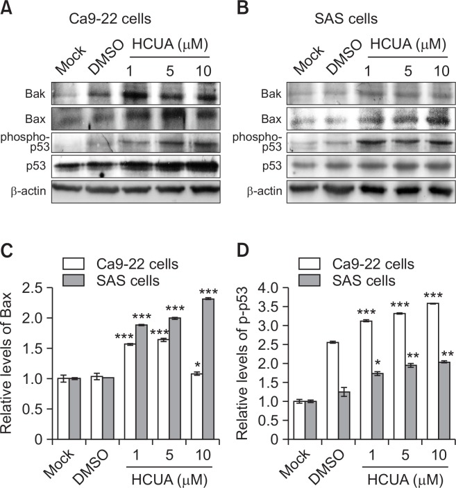
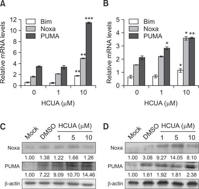
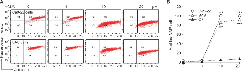
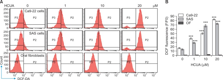
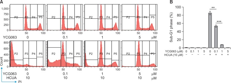
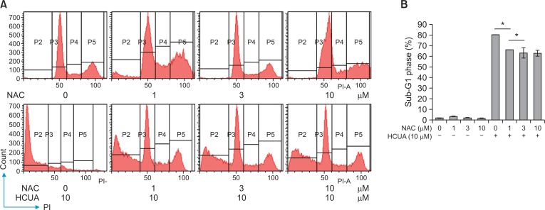
Cis-3-O-p-hydroxycinnamoyl ursolic acid (HCUA), a triterpenoid compound, was purified from Elaeagnus oldhamii Maxim. This traditional medicinal plant has been used for treating rheumatoid arthritis and lung disorders as well as for its anti-inflammation and anticancer activities. This study aimed to investigate the anti-proliferative and apoptotic-inducing activities of HCUA in oral cancer cells. HCUA exhibited anti-proliferative activity in oral cancer cell lines (Ca9-22 and SAS cells), but not in normal oral fibroblasts. The inhibitory concentration of HCUA that resulted in 50% viability was 24.0 µM and 17.8 µM for Ca9-22 and SAS cells, respectively. Moreover, HCUA increased the number of cells in the sub-G1 arrest phase and apoptosis in a concentration-dependent manner in both oral cancer cell lines, but not in normal oral fibroblasts. Importantly, HCUA induced p53-mediated transcriptional regulation of pro-apoptotic proteins (Bax, Bak, Bim, Noxa, and PUMA), which are associated with mitochondrial apoptosis in oral cancer cells via the loss of mitochondrial membrane potential. HCUA triggered the production of intracellular reactive oxygen species (ROS) that was ascertained to be involved in HCUA-induced apoptosis by the ROS inhibitors YCG063 and N-acetyl-L-cysteine. As a result, HCUA had potential antitumor activity to oral cancer cells through eliciting ROS-dependent and p53-mediated mitochondrial apoptosis. Overall, HCUA could be applicable for the development of anticancer agents against human oral cancer.

Keywords: Cis-3-O-p-hydroxycinnamoyl ursolic acid; Mitochondrial apoptosis; Oral cancer cells; ROS.


Conflict of interest statement

CONFLICT OF INTEREST

All authors had no conflict of interest.

Figures

Fig. 1.
Fig. 1.
Survival rates of oral cancer cell lines (Ca9-22 and SAS) and normal oral fibroblasts (OF) in response to cis-3-O-p-hydroxy-cinnamoyl ursolic acid (HCUA). The structure of HCUA is shown in (A). Ca9-22, SAS, and OF cells were treated with HCUA for 48 h, and then the survival rate of treated cells was calculated based on MTT assays (B). **p<0.01, ***p<0.001 compared with mock cells.
Fig. 2.
Fig. 2.
Effect of cis-3-O-p-hydroxycinnamoyl ursolic acid (HCUA) on cell cycle profile of oral cancer cell lines (Ca9-22 and SAS) and normal oral fibroblasts (OF). Cells were harvested 48 h post-treatment, stained with propidium iodide, and then analyzed by flow cytometry (A). The percent of Ca9-22, SAS, and OF cells in the sub G1 (apoptotic) phase after treatment with HCUA was determined (B). **p<0.01, ***p<0.001 compared with mock cells.
Fig. 3.
Fig. 3.
Induction of apoptosis in oral cancer cells by cis-3-O-p-hydroxycinnamoyl ursolic acid (HCUA). Ca9-22 and SAS cells as well as oral fibroblasts were harvested 48 h post-treatment, double-stained with Annexin V-FITC/PI, and then analyzed by flow cytometry (A). The percentage of cells that were Annexin V-positive/PI-negative (early phase of apoptosis) is shown (B). The percentage of cells that were Annexin V-positive/PI-positive (late apoptosis) is shown (C). *p <0.05, **p <0.01, ***p<0.001 compared with mock cells.
Fig. 4.
Fig. 4.
Relative protein of pro-apoptotic genes in cis-3-O-p-hydroxy-cinnamoyl ursolic acid (HCUA)-treated cells. The protein levels of Bak, Bax, phospho-p53 (Ser15), and p53 in Ca9-22 (A) and SAS (B) cells treated with HCUA were characterized 48 h post-treatment by western blot analysis. The intensity of each immuno-reactive band for Bax or phospho-p53 (Ser15) was quantified using Image J; their relative intensities were normalized by β-actin (C, D), respectively. *p<0.05, **p<0.01, ***p<0.001 compared with mock cells.
Fig. 5.
Fig. 5.
Relative mRNA and protein levels of p53-mediated transcriptional genes in cis-3-O-p-hydroxycinnamoyl ursolic acid (HCUA)-treated cells. Relative mRNA and protein levels of Bim, Noxa, and PUMA in HCUA-treated Ca9-22 (A, C) and of SAS (B, D) cells were measured using real-time RT-PCR and western blotting, respectively. Their relative levels were normalized by GAPDH mRNA or β-actin protein. *p<0.05, **p<0.01, ***p<0.001 compared with mock cells.
Fig. 6.
Fig. 6.
Loss of mitochondrial membrane potential (ΔΨM) in Ca9-22 and SAS cells treated with cis-3-O-p-hydroxycinnamoyl ursolic acid (HCUA). Oral cancer cells and fibroblasts were treated with HCUA and then harvested at 48 h post-treatment, stained with DiOC6(3), and then analyzed by flow cytometry (A). The relative changes in the percentage of low MMP cells in response to HCUA are shown (B). ***p<0.001 compared with mock cells.
Fig. 7.
Fig. 7.
Increase of intracellular ROS production in cis-3-O-p-hydroxycinnamoyl ursolic acid (HCUA)-treated cells. Ca9-22, SAS, and OF cells were harvested 48 h post-treatment with HCUA, stained with 2,7-dichlorodihydrofluorescein diacetate (DCF-DA), and then analyzed by flow cytometry (A). Relative fluorescent intensity of DCF in treated-cells was subsequently measured (B). ***p<0.001 compared with mock cells.
Fig. 8.
Fig. 8.
Reduction of cis-3-O-p-hydroxycinnamoyl ursolic acid (HCUA)-induced apoptosis in Ca9-22 cells by YCG063. Cells were treated with YCG063, HCUA, or a combination of both, harvested 48 h post-treatment, fixed by 70% ethanol, stained with a propidium iodide solution, and then examined by flow cytometry (A). The percentage of cells in the sub-G1 (apoptotic) phase is shown in (B). **p<0.01, ***p<0.001.
Fig. 9.
Fig. 9.
Inhibitory effect of NAC on cis-3-O-p-hydroxycinnamoyl ursolic acid (HCUA)-induced apoptosis. Ca9-22 cells were treated with NAC, HCUA, or a combination of both, harvested 48 h post-treatment, stained with propidium iodide, and then examined by flow cytometry (A). The percentage of cells in the sub-G1 (apoptotic) phase is shown in (B). *p<0.05.

Similar articles

Cited by

References

    1. Cai Q, Lin J, Zhang L, Lin J, Wang L, Chen D, Peng J. Comparative proteomics-network analysis of proteins responsible for ursolic acid-induced cytotoxicity in colorectal cancer cells. Tumour. Biol. 2017;39:1010428317695015. doi: 10.1177/1010428317695015. - DOI - PubMed
    1. Chang CF, Kuo YL, Pu C, Chou Y. J. Neck dissection and stroke in patients with oral cavity cancer: a population-based cohort study. Head Neck. 2016;39:63–70. doi: 10.1002/hed.24535. - DOI - PubMed
    1. Chen J, Fu H, Wang Z, Yin F, Li J, Hua Y, Cai Z. A new synthetic ursolic acid derivative IUA with anti-tumor efficacy against osteosarcoma cells via inhibition of JNK signaling pathway. Cell Physiol Biochem. 2014;34:724–733. doi: 10.1159/000363037. - DOI - PubMed
    1. Chipuk JE, Moldoveanu T, Llambi F, Parsons MJ, Green DR. The BCL-2 family reunion. MolCell. 2010;37:299–310. - PMC - PubMed
    1. Dar BA, Lone AM, Shah WA, Qurishi MA. Synthesis and screening of ursolic acid-benzylidine derivatives as potential anti-cancer agents. Eur. J. Med. Chem. 2016;111:26–32. doi: 10.1016/j.ejmech.2016.01.026. - DOI - PubMed

LinkOut - more resources