. 2018 Sep 27;15(1):282.
doi: 10.1186/s12974-018-1319-x.

α1-antitrypsin mitigates NLRP3-inflammasome activation in amyloid β1-42-stimulated murine astrocytes

Affiliations

α1-antitrypsin mitigates NLRP3-inflammasome activation in amyloid β1-42-stimulated murine astrocytes

Taraneh Ebrahimi et al. J Neuroinflammation. .

Abstract

Background: Neuroinflammation has an essential impact on the pathogenesis and progression of Alzheimer's disease (AD). Mostly mediated by microglia and astrocytes, inflammatory processes lead to degeneration of neuronal cells. The NLRP3-inflammasome (NOD-like receptor family, pyrin domain containing 3) is a key component of the innate immune system and its activation results in secretion of the proinflammatory effectors interleukin-1β (IL-1β) and interleukin-18 (IL-18). Under physiological conditions, cytosolic NLRP3-inflammsome is maintained in an inactive form, not able to oligomerize. Amyloid β1-42 (Aβ1-42) triggers activation of NLRP3-inflammasome in microglia and astrocytes, inducing oligomerization and thus recruitment of proinflammatory proteases. NLRP3-inflammasome was found highly expressed in human brains diagnosed with AD. Moreover, NLRP3-deficient mice carrying mutations associated with familial AD were partially protected from deficits associated with AD. The endogenous protease inhibitor α1-antitrypsin (A1AT) is known for its anti-inflammatory and anti-apoptotic properties and thus could serve as therapeutic agent for NLRP3-inhibition. A1AT protects neurons from glutamate-induced toxicity and reduces Aβ1-42-induced inflammation in microglial cells. In this study, we investigated the effect of Aβ1-42-induced NLRP3-inflammasome upregulation in primary murine astrocytes and its regulation by A1AT.

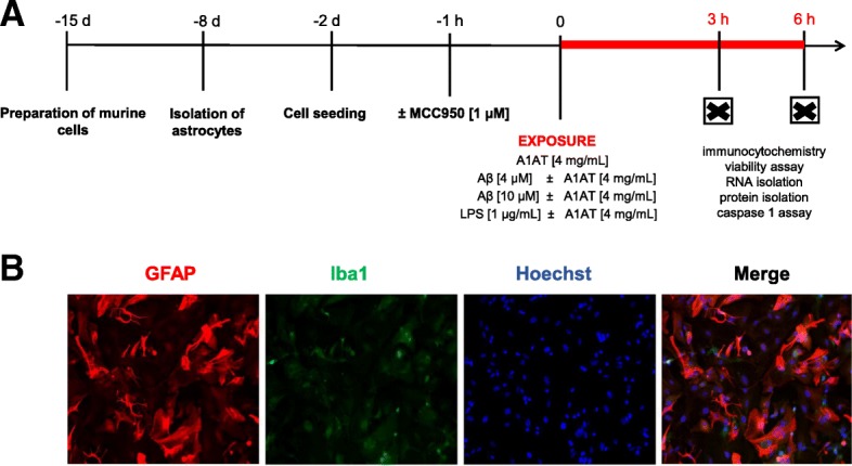
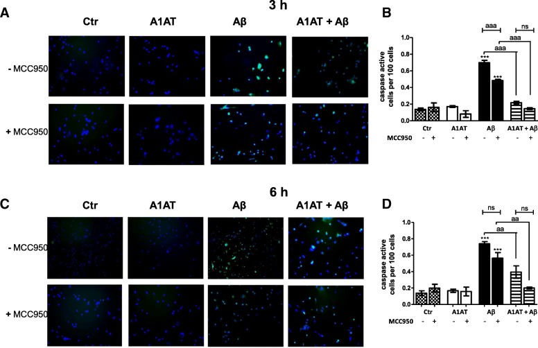
Methods: Primary cortical astrocytes from BALB/c mice were stimulated with Aβ1-42 and treated with A1AT. Regulation of NLRP3-inflammasome was examined by immunocytochemistry, PCR, western blot and ELISA. Our studies included an inhibitor of NLRP3 to elucidate direct interactions between A1AT and NLRP3-inflammasome components.

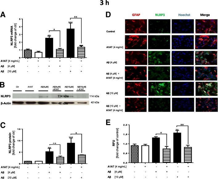
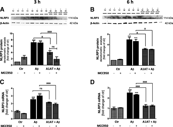
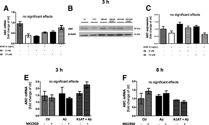
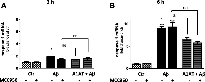
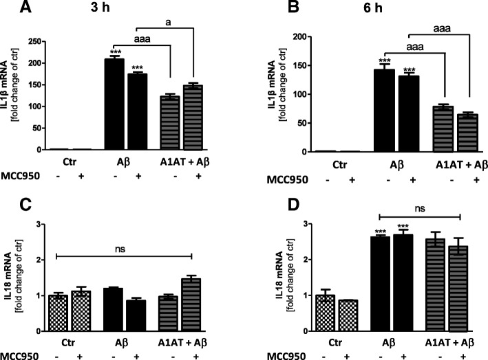
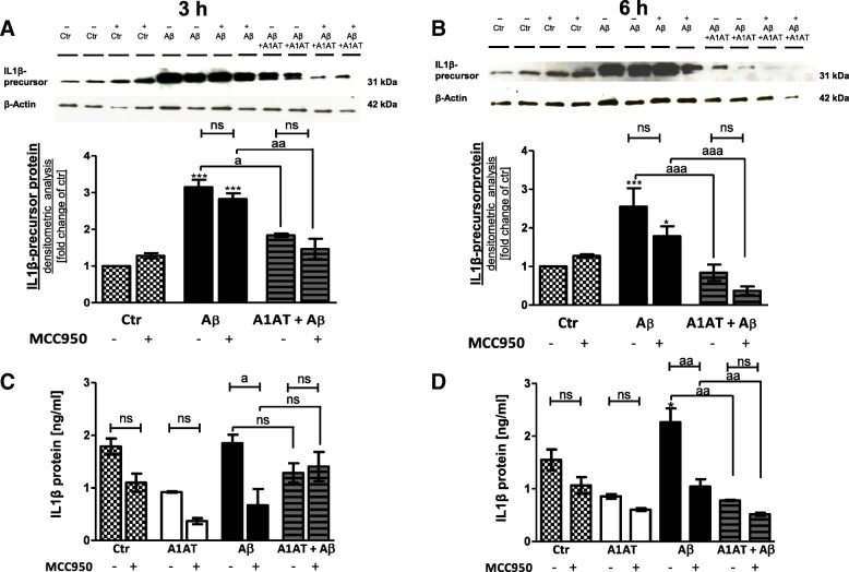
Results: Our study revealed that A1AT reduces Aβ1-42-dependent upregulation of NLRP3 at the mRNA and protein levels. Furthermore, A1AT time-dependently mitigated the expression of caspase 1 and its cleavage product IL-1β in Aβ1-42-stimulated astrocytes.

Conclusion: We conclude that Aβ1-42-stimulation results in an upregulation of NLRP3, caspase 1, and its cleavage products in astrocytes. A1AT time-dependently hampers neuroinflammation by downregulation of Aβ1-42-mediated NLRP3-inflammasome expression and thus may serve as a pharmaceutical opportunity for the treatment of Alzheimer's disease.

Keywords: Alpha 1-antitrypsin; Alzheimer’s disease; Amyloid β; Astrocytes; Inflammasome; NALP3; NLRP3; Neuroinflammation.


Conflict of interest statement

Ethics approval

No human tissue was involved in this study. Postnatal (P0 to P2) cortical astrocyte culture preparation from BALB/c mice (Charles River) was performed as previously described by Habib et al. 2014 [51]. Preparation was conducted in accordance with animal welfare policy of University Hospital Aachen and the government of the State of North Rhine-Westphalia, Germany (no. 84.02.04.2015.A292).

Consent for publication

Not applicable

Competing interests

All authors declare that they have no competing interests.

Publisher’s Note

Springer Nature remains neutral with regard to jurisdictional claims in published maps and institutional affiliations.

Figures

Fig. 1
Fig. 1
Experimental setting. a Cortices from BALB/c mice were prepared and seeded on flasks 2 weeks prior to stimulation. After a week, when cell confluence was about 80%, flasks were shaken to remove microglia and oligodendrocytes from astrocytes. At passage 2, astrocytes were seeded on experimental plates 48 h prior to stimulation. After short-term stimulation (3 h/ 6 h) with two concentrations of Aβ1–42 (4, 10 μM) or LPS (1 μg/mL) and treatment with A1AT (4 mg/mL) immunocytochemistry, viability assays, caspase 1 assay, RNA and protein isolation were performed. For our control experiments, pretreatment with MCC950 (1 μM) was performed 1 h prior to stimulation. b Astrocyte purity was assessed by ICC using GFAP and Iba1 combined with Hoechst for DNA staining
Fig. 2
Fig. 2
Effect of Aβ1–42 stimulation and A1AT treatment ± MCC950 on cell viability at 3 h and 6 h. Treatment with Aβ1–42 [4 μM], A1AT [4 mg/mL], and MCC950 [1 μM] did not affect LDH-release at 3 h and 6 h stimulation, whereas LPS-stimulation [1 μg/mL] significantly increased LDH-release (a, c). Cell lysis defined maximal (100%; 1.0) release of LDH. MTT-assay (b, d) revealed a trend towards decrease in cell metabolism after 3 h and 6 h Aβ1–42-stimulation, which was not significant. Treatment with MCC950 led to an increase of cell metabolism in each treatment group at 3 h and 6 h, which was not significant. */ap < 0.05; **/aap < 0.01; ***/aaap < 0.001, ns not significant compared to untreated cell control (ctr)
Fig. 3
Fig. 3
Treatment with A1AT abrogated Aβ1–42-induced upregulation of NLRP3 mRNA and protein in primary astrocytes. a As quantified by RT-PCR treatment with 4 μM and 10 μM of Aβ1–42 significantly increased mRNA levels of NLRP3 in astrocytes. In addition, co-treatment with 4 mg/mL of A1AT blocked this increase in NLRP3 mRNA expression significantly. Data of n = 6 in triplicate represent mean ± SD. bc Densitometric analysis of western blots confirmed an Aβ1–42-induced significant increase of NLRP3 protein levels at 3 h. Treatment with 4 mg/mL of A1AT significantly attenuated this increase at 10 μM Aβ1–42. Data of n = 3 in triplicate represent mean ± SD. de Astrocytes treated with 10 μM of Aβ1–42 showed a significant increase of NLRP3-fluorescence intensity, whereas co-treatment with A1AT significantly reduced NLRP3-expression. Fluorescence images of each experiment were taken using the exact same microscope settings and fluorescence intensity was measured by ImageJ (USA). Data of n = 4 in triplicate represent mean ± SD. */ap < 0.05; **/aap < 0.01; ***/aaap < 0.001, ns not significant compared to untreated cell control (ctr)
Fig. 4
Fig. 4
Treatment with A1AT abrogated Aβ1–42-induced upregulation of NLRP3 mRNA and protein in primary astrocytes. Western blot confirmed an Aβ1–42-induced significant increase of NLRP3 protein levels at 3 h (a) and 6 h (b). Co-treatment with A1AT significantly attenuated this increase at 3 h (a) and 6 h (b) stimulation time. Addition of MCC950 did not alter NLRP3-protein levels in all treatment groups. Treatment with Aβ1–42 significantly increased mRNA levels of NLRP3 in astrocytes at 3 h (c) and 6 h (d). Co-treatment with A1AT significantly blocked this increase in NLRP3 mRNA expression. MCC950-pretreatment had no effects on NLRP3 mRNA expression at 3 h and 6 h. Data of n = 3 in triplicate represent mean ± SD. */ap < 0.05; **/aap < 0.01; ***/aaap < 0.001, ns not significant compared to untreated cell control (ctr)
Fig. 5
Fig. 5
ASC was not regulated by Aβ1–42 and A1AT at 3 h and 6 h stimulation. (a) Quantitative RT-PCR demonstrated ASC was not regulated by Aβ1–42 or A1AT. Data of n = 6 in triplicate represent mean ± SD. (bc) Western blot revealed no regulation of ASC by Aβ1–42 or A1AT. Data of n = 3 in triplicate represent mean ± SD. (e–f) RT-PCR demonstrated that also at 6 h stimulation time such as MCC950-pretreatment did not regulate ASC mRNA expression. Data of n = 3 in triplicate represent mean ± SD. */ap < 0.05; **/aap < 0.01; ***/aaap < 0.001, ns not significant compared to untreated cell control (ctr)
Fig. 6
Fig. 6
A1AT time-dependently blocked Aβ1–42-induced upregulation of caspase 1 mRNA in primary astrocytes. (b–c) As quantified by RT-PCR treatment with 4 μM Aβ1–42 significantly increased mRNA levels of caspase 1 in astrocytes at 6 h, but not at 3 h. Co-treatment with A1AT blocked this increase in caspase 1 mRNA expression significantly at 6 h, but not at 3 h. Addition of MCC950 had no effects on caspase 1 mRNA expression at both stimulation times. Data of n = 3 in triplicate represent mean ± SD.*/ap < 0.05; **/aap < 0.01; ***/aaap < 0.001, ns not significant compared to untreated cell control (ctr)
Fig. 7
Fig. 7
A1AT mitigated caspase activity in Aβ1–42-stimulated astrocytes. (a, c) For convenience, we have only illustrated the overlay images of all treatment groups ± MCC950. Counting of caspase active cells (b, d) revealed a significant increase of caspase active cells with 4 μM Aβ1–42-stimulation at both stimulation times. Co-treatment with A1AT significantly blocked this effect at both stimulation times. In MCC950-treated and Aβ1–42-stimulated cells caspase activity significantly declined at 3 h. Additive treatment by MCC950 to A1AT and Aβ1–42-stimulated astrocytes did not significantly change caspase activity. Data of n = 3 in triplicate represent mean ± SD. */ap < 0.05; **/aap < 0.01; ***/aaap < 0.001, ns not significant compared to untreated cell control (ctr)
Fig. 8
Fig. 8
A1AT mitigated Aβ1–42-induced upregulation of IL-1β mRNA in primary astrocytes. (ab) As quantified by RT-PCR stimulation with 4 μM Aβ1–42 significantly increased mRNA levels of IL-1β in astrocytes at 3 h and 6 h. Co-treatment with A1AT blocked this increase significantly at 3 h and 6 h. Addition of MCC950 had no significant impact on gene expression at 3 h and 6 h. (c) Aβ1–42-stimulation such as A1AT-treatment did not affect IL-18 mRNA expression at 3 h. (d) Stimulation with Aβ1–42 significantly increased mRNA levels of IL-18 in astrocytes at 6 h. Co-treatment with A1AT did not block this increase. MCC950 had no effects on gene expression of IL-18 at 3 h and 6 h. Data of n = 3 in triplicate represent mean ± SD. */ap < 0.05; **/aap < 0.01; ***/aaap < 0.001, ns not significant compared to untreated cell control (ctr)
Fig. 9
Fig. 9
Treatment with A1AT time-dependently mitigated Aβ1–42-induced upregulation of IL1β-precursor protein in primary astrocytes. Western blot confirmed an Aβ1–42-induced significant increase of of IL1β-precursor protein at 3 h (a) and 6 h (b). Co-treatment with A1AT significantly attenuated this increase at 3 h and 6 h stimulation time. Addition of MCC950 did not alter levels of IL1β-precursor protein in Aβ1–42-stimulated astrocytes. A1AT + in Aβ1–42 in the presence of MCC950 did not alter IL1β-precursor protein levels at both 3 h and 6 h. (cd) IL-1β-ELISA revealed a time-dependent upregulation of IL-1β protein levels in Aβ1–42-stimulated astrocytes, significant at 6 h stimulation. Co-treatment with A1AT significantly decreased protein expression of IL-1β time-dependently, at 6 h stimulation time. The presence of MCC950 in Aβ1–42-stimulated decreased IL-1β-expression significantly at both 3 h and 6 h. Further, there is a trend towards a decrease of IL-1β-protein levels in the presence of MCC950 in nearly all treatment groups, though not significant. Co-stimulation of Aβ1–42 and A1AT in MCC950-pretreated astrocytes did not affect IL-1β-expression. Data of n = 3 in triplicate represent mean ± SD. */ap < 0.05; **/aap < 0.01; ***/aaap < 0.001, ns not significant compared to untreated cell control (ctr)

References

    1. Prince M, Bryce R, Albanese E, Wimo A, Ribeiro W, Ferri CP. The global prevalence of dementia: a systematic review and metaanalysis. Alzheimers Dement. 2013;9(1):63–75.e62. doi: 10.1016/j.jalz.2012.11.007. - DOI - PubMed
    1. Brookmeyer R, Johnson E, Ziegler-Graham K, Arrighi HM. Forecasting the global burden of Alzheimer's disease. Alzheimers Dement. 2007;3(3):186–191. doi: 10.1016/j.jalz.2007.04.381. - DOI - PubMed
    1. Hirtz D, Thurman DJ, Gwinn-Hardy K, Mohamed M, Chaudhuri AR, Zalutsky R. How common are the “common” neurologic disorders? Neurology. 2007;68(5):326–337. doi: 10.1212/01.wnl.0000252807.38124.a3. - DOI - PubMed
    1. Rosenberg PB, Lyketsos C. Mild cognitive impairment: searching for the prodrome of Alzheimer's disease. World Psychiatry. 2008;7(2):72–78. doi: 10.1002/j.2051-5545.2008.tb00159.x. - DOI - PMC - PubMed
    1. Wilson RS, Leurgans SE, Boyle PA, Bennett DA. Cognitive decline in prodromal Alzheimer disease and mild cognitive impairment. Arch Neurol. 2011;68(3):351–356. doi: 10.1001/archneurol.2011.31. - DOI - PMC - PubMed

MeSH terms

Substances