. 2018 Sep 28;361(6409):eaao4227.
doi: 10.1126/science.aao4227.

Neutrophil extracellular traps produced during inflammation awaken dormant cancer cells in mice

Affiliations

Neutrophil extracellular traps produced during inflammation awaken dormant cancer cells in mice

Jean Albrengues et al. Science. .

Abstract

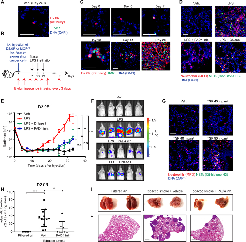
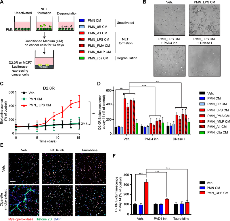
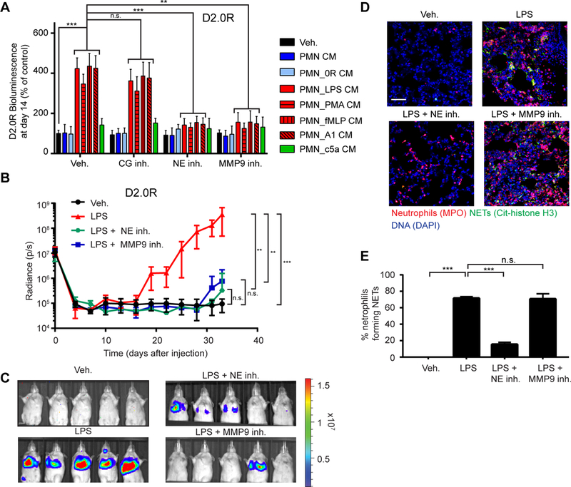
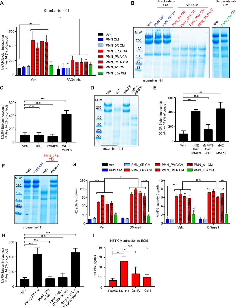
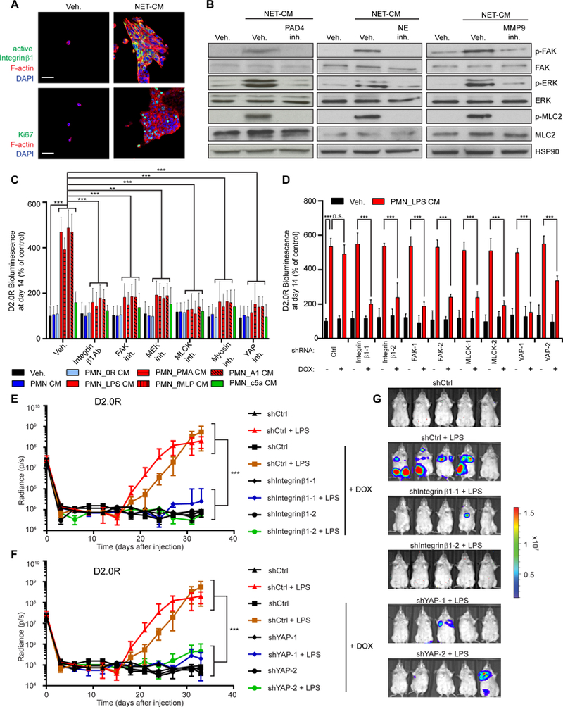
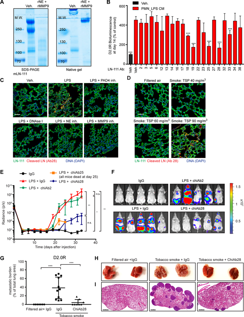
Cancer cells from a primary tumor can disseminate to other tissues, remaining dormant and clinically undetectable for many years. Little is known about the cues that cause these dormant cells to awaken, resume proliferating, and develop into metastases. Studying mouse models, we found that sustained lung inflammation caused by tobacco smoke exposure or nasal instillation of lipopolysaccharide converted disseminated, dormant cancer cells to aggressively growing metastases. Sustained inflammation induced the formation of neutrophil extracellular traps (NETs), and these were required for awakening dormant cancer. Mechanistic analysis revealed that two NET-associated proteases, neutrophil elastase and matrix metalloproteinase 9, sequentially cleaved laminin. The proteolytically remodeled laminin induced proliferation of dormant cancer cells by activating integrin α3β1 signaling. Antibodies against NET-remodeled laminin prevented awakening of dormant cells. Therapies aimed at preventing dormant cell awakening could potentially prolong the survival of cancer patients.


Figures

Fig. 1.
Fig. 1.. NETs promote dormant cancer cell awakening after sustained lung inflammation.
(A, B) D2.0R cells stayed dormant in the lungs of mice for months. (A) Representative micrographs of D2.0R cells (red, mCherry) in Ki67 (green) and DAPI (blue) stained lung sections from untreated mice at day 240. Scale bar: 50 μm. (B) Schematic showing experimental design. (C) Sustained lung inflammation promoted awakening. Representative immunostaining of lungs for D2.0R cells (red, mCherry) and Ki67 (green) with DAPI (blue), at indicated time after injection. Scale bars: 50 μm. (D) Nasal LPS instillation induced NETs in lungs. Representative immunostaining for myeloperoxidase (MPO, red), citrullinated histone H3 (Cit-histone H3, green) and DAPI (blue) in the lungs of mice treated as indicated. Scale bar: 50 μm. (E, F) Targeting NETs reduced inflammation-induced awakening. (E) Mice with D2.0R cells, treated as indicated, were monitored by bioluminescence imaging (BLI) (n=10 mice for vehicle and LPS groups; n=5 mice for DNase I and PAD4 inhibitor groups; mean±SD). (F) Representative BLI images at day 33. (G) Exposure to tobacco smoke (TSP for total suspended particles) induced NETs in the lungs. Representative immunostaining for myeloperoxidase (red), citrullinated histone H3 (green) and DAPI (blue) in the lungs of mice treated as indicated. Scale bar: 50 μm. (H-J) Targeting NETs reduced smoking-induced awakening. (H) Mice with D2.0R cells were treated as indicated, and lung metastatic burden quantified from hematoxylin and eosin (H&E) staining (n=8 mice for filtered air and tobacco smoke + PAD4 groups; n=12 mice for tobacco smoke + vehicle group; mean±SD). (I) Lungs from mice treated as indicated at day 30. (J) Representative images of H&E staining of lungs from panel (H) at day 30. Scale bar: 700 μm.
Fig. 2.
Fig. 2.. NETs induce awakening in vitro in the absence of other host cells.
(A) Experimental design for inducing NETs or degranulation neutrophils (PMNs). Conditioned medium (CM) was collected 20 hours after activation and added to cultures of luciferase-expressing cancer cells plated on top of matrigel. (B-D) NET-containing CM induced awakening of D2.0R cells on matrigel. (B) Representative images of 3D cultures at day 15, treated as indicated. Scale bar: 250 μm. (C) BLI quantification over 15 days with indicated treatments (n=3; mean±SD). (D) BLI signal 14 days after indicated treatments. PAD4 inhibitor and DNase I were used during neutrophil culture to block and digest NET formation, respectively (n=3; mean±SD). (E, F) Cigarette smoke extract (CSE) induced the formation of NETs and subsequent awakening in vitro. (E) Immunostaining of mouse neutrophils cultured as indicated. DAPI (blue), anti-MPO (red), and anti-histone H2B (green) staining were used to assess NET formation. Scale bar: 100 μm. (F) BLI signal 14 days after indicated treatments. Taurolidine and PAD4 inhibitor were used during neutrophil culture to block NET formation (n=3; mean±SD).
Fig. 3.
Fig. 3.. NET-associated NE and MMP9 induce awakening from dormancy through extracellular matrix remodeling.
(A) NE and MMP9 were required for NET-induced awakening of D2.0R cells in vitro. BLI signal of luciferase-expressing cells 14 days after indicated treatments. CG, NE, and MMP9 inhibitors were used during cancer cell culture (n=3; mean±SD). (B, C) NE and MMP9 activity were required for LPS-induced awakening in vivo. (B) Mice with D2.0R cells were treated as indicated and monitored by BLI (n=10 mice for vehicle and LPS groups; n=5 mice for NE inhibitor and MMP9 inhibitor groups; mean±SD). (C) Representative BLI images at day 33. (D, E) NE, but not MMP9 inhibition, prevented formation of NETs after LPS-induced lung inflammation. (D) DAPI (blue), anti-MPO (red), and anti-citrullinated histone H3 (green) staining were used to assess NET formation in the lungs of mice treated as indicated. Scale bar: 50 μm. (E) Quantification of NET-forming neutrophils (n=3 mice; mean±SD).
Fig. 4.
Fig. 4.. NETs facilitate laminin-111 remodeling.
(A) Culturing cancer cells on laminin-111 allowed NET-dependent control of dormancy and awakening. BLI signal of luciferase-expressing D2.0R cells after 14 days under indicated conditions (PAD4 inhibitor was used during PMN culture, n=3; mean±SD). (B) NET-containing CM cleaved laminin-111. Cleavage of laminin-111 after incubation with indicated CM was detected by SDS-PAGE, under reducing and denaturing conditions, and coomassie blue staining. (C-E) Both NE and MMP9 were required for awakening and laminin-111 cleavage. Laminin-111 was incubated with recombinant proteases alone (C) or added sequentially (E), and D2.0R cells were cultured on the remodeled matrix (C, E) or laminin-111 was analyzed by SDS-PAGE (D). BLI was used to quantify the cancer cells after 14 days (for experimental design, see Fig. S22B) (n=3; mean±SD). (F) NET-associated proteases had higher laminin-111 cleavage activity than DNase-released NE and MMP9. Laminin-111 was incubated as indicated before SDS-PAGE. DNase I was added after PMN culture. (G) NE and MMP activity toward soluble fluorescent substrates was not reduced by DNase I treatment of NET-containing CM. NE and MMP activity in the indicated PMN CM. DNase I was used after PMN activation (n=3; mean±SD). (H) NET-associated proteases had higher awakening activity than free NE and MMP9. Laminin-111 was incubated as indicated before D2.0R cell culture. BLI quantification of cancer cells at day 14 (n=3; mean±SD). (I) NETs bound preferentially to laminin-111. CM from neutrophils stimulated with LPS to form NETs was incubated on plastic or ECM-coated plates as indicated. dsDNA bound to ECM proteins were quantified (n=3; mean±SD).
Fig. 5.
Fig. 5.. Integrin β1 signaling pathway regulates NET-induced awakening.
(A) NETs activated integrin β1 and proliferation in dormant cancer cells. D2.0R cells cultured on matrigel-coated 0.2 kPa hydrogel stained for active integrin β1 (green, top) or Ki67 (green, bottom), phalloidin (red), and DAPI (blue). Scale bar: 50 μm. (B) NET-containing CM activated the integrin β1 signaling pathway. D2.0R cells cultured on matrigel for 10 days under indicated conditions, analyzed for phospho-FAK, phospho-ERK½, and phospho-MLC2 by Western blot. Controls: total FAK, ERK½, MLC2, and Heat Shock Protein 90 (HSP90). (C, D) Activation of the integrin β1 pathway is required for NET-induced awakening. (C) BLI of luciferase-expressing D2.0R cells cultured on matrigel for 14 days under indicated conditions (n=3; mean±SD), and (D) cultured on matrigel using doxycycline to induce expression of indicated shRNAs (n=3; mean±SD). (E-G) Inhibition of integrin-mediated YAP activation prevents LPS-induced awakening. (E, F) Mice with inducible shRNA-expressing D2.0R cells were monitored by BLI. Doxycycline treatment was started on day 5 (n=5 mice per group; mean±SD). (G) Representative BLI images at day 33.
Fig. 6.
Fig. 6.. A NET-generated laminin epitope promotes awakening.
(A) Laminin-111 was not fully degraded by NET-associated proteases. Laminin-111 was incubated with rNE and rMMP9, and cleavage was assessed by SDS-PAGE and coomassie blue staining under reducing/denaturing or non-reducing/non-denaturing conditions. (B) Antibodies against NE- and MMP9-cleaved laminin-111 prevented NET-mediated awakening. BLI of luciferase-expressing D2.0R cells cultured on laminin-111, 14 days after treatment with indicated antibody clone (n=3; mean±SD). (C, D) Lung inflammation and NETs induce production of cleaved laminin. Immunostaining of lungs for DAPI (blue), full-length laminin-111 (ab11575 from Abcam, green), and cleaved laminin antibody 28 (red), treated as indicated. Total suspended particles (TSP) in cigarette smoke exposure conditions indicated. Scale bars: 50 μm. (E-I) Antibodies against NET-remodeled laminin prevented inflammation-induced awakening in vivo. (E) Mice with D2.0R cells were treated as indicated and monitored by BLI (n=5 mice per group; mean±SD). (F) Representative BLI images at day 33. (G) Metastatic burden in lungs from mice exposed to tobacco smoke quantified with H&E staining. (H) Representative photos of lung. (I) Representative H&E images of lungs from panel (H) at day 30 (n=7 mice for filtered air + IgG, n=8 mice for tobacco smoke + ChiAb28 inhibitor, n=11 mice for tobacco smoke + IgG; mean±SD). Scale bar: 700 μm.

Comment in

  • How dormant cancer persists and reawakens.
    Aguirre-Ghiso JA. Aguirre-Ghiso JA. Science. 2018 Sep 28;361(6409):1314-1315. doi: 10.1126/science.aav0191. Science. 2018. PMID: 30262486 Free PMC article. No abstract available.
  • NETs awaken sleeping cancer cells.
    Bordon Y. Bordon Y. Nat Rev Immunol. 2018 Nov;18(11):665. doi: 10.1038/s41577-018-0081-8. Nat Rev Immunol. 2018. PMID: 30327547 No abstract available.
  • Waking up in a trap.
    Dart A. Dart A. Nat Rev Cancer. 2018 Dec;18(12):722-723. doi: 10.1038/s41568-018-0071-y. Nat Rev Cancer. 2018. PMID: 30361637 No abstract available.
  • Mets and NETs: The Awakening Force.
    Sanz-Moreno V, Balkwill FR. Sanz-Moreno V, et al. Immunity. 2018 Nov 20;49(5):798-800. doi: 10.1016/j.immuni.2018.11.009. Immunity. 2018. PMID: 30462996

References

    1. Sosa MS, Bragado P,Aguirre-Ghiso JA, Mechanisms of disseminated cancer cell dormancy: an awakening field. Nat Rev Cancer 14, 611–622 (2014). - PMC - PubMed
    1. Malladi S. et al., Metastatic latency and immune evasion through autocrine inhibition of WNT. Cell 165, 45–60 (2016). - PMC - PubMed
    1. Romero I,Garrido F,Garcia-Lora AM, Metastases in immune-mediated dormancy: a new opportunity for targeting cancer. Cancer Res 74, 6750–6757 (2014). - PubMed
    1. Farrar JD et al., Cancer dormancy. VII. A regulatory role for CD8+ T cells and IFN-gamma in establishing and maintaining the tumor-dormant state. J Immunol 162, 2842–2849 (1999). - PubMed
    1. Muller M. et al., EblacZ tumor dormancy in bone marrow and lymph nodes: active control of proliferating tumor cells by CD8+ immune T cells. Cancer Res 58, 5439–5446 (1998). - PubMed

Publication types

MeSH terms