. 2018 Sep 27;8(1):14440.
doi: 10.1038/s41598-018-32748-w.

Bioprocessing strategies to enhance the challenging isolation of neuro-regenerative cells from olfactory mucosa

Affiliations

Bioprocessing strategies to enhance the challenging isolation of neuro-regenerative cells from olfactory mucosa

Melanie Georgiou et al. Sci Rep. .

Abstract

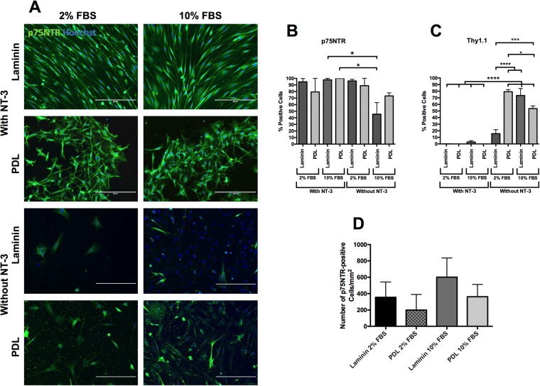
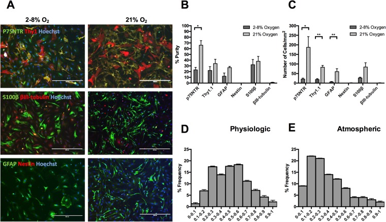
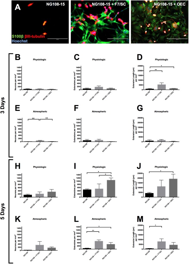
Olfactory ensheathing cells (OECs) are a promising potential cell therapy to aid regeneration. However, there are significant challenges in isolating and characterizing them. In the current study, we have explored methods to enhance the recovery of cells expressing OEC marker p75NTR from rat mucosa. With the addition of a 24-hour differential adhesion step, the expression of p75NTR was significantly increased to 73 ± 5% and 46 ± 18% on PDL and laminin matrices respectively. Additionally, the introduction of neurotrophic factor NT-3 and the decrease in serum concentration to 2% FBS resulted in enrichment of OECs, with p75NTR at nearly 100% (100 ± 0% and 98 ± 2% on PDL and laminin respectively), and candidate fibroblast marker Thy1.1 decreased to zero. Culturing OECs at physiologically relevant oxygen tension (2-8%) had a negative impact on p75NTR expression and overall cell survival. Regarding cell potency, co-culture of OECs with NG108-15 neurons resulted in more neuronal growth and potential migration at atmospheric oxygen. Moreover, OECs behaved similarly to a Schwann cell line positive control. In conclusion, this work identified key bioprocessing fundamentals that will underpin future development of OEC-based cell therapies for potential use in spinal cord injury repair. However, there is still much work to do to create optimized isolation methods.


Conflict of interest statement

The authors declare no competing interests.

Figures

Figure 1
Figure 1
OEC isolation and culture from adult Sprague-Dawley female rats. This figure summarizes the best identified method of isolating OECs from rat primary tissue. It includes the initial dissection, the enzymatic digestion, the differential adhesion step as well as the full 14 days of the OEC culture (TCP = tissue culture plastic).
Figure 2
Figure 2
Differential Adhesion Step: Fluorescent micrographs of mucosa-derived OECs after the 24-hour differential adhesion on PDL (A), and Laminin (B). Scale bars are 100 µm. (C) Fluorescent micrographs of remaining adherent cells in the differential adhesion step. Following the cell isolation process, mucosa-derived cells were seeded either directly onto PDL or laminin for a 2-week culture period; or were first seeded on to TCP for 24 hours, before the supernatant was re-seeded onto PDL or laminin for the remainder of the 2-week culture period. All cells were cultured in DMEM/F12 + 1% P/S + 10% FBS. Mucosa-derived cells were fixed overnight at day 14 and stained to detect p75NTR protein. From these images, it can be seen that no glial cells were left behind due to the lack of positive glial cell staining (p75NTR, S100β) and the presence of fibroblastic markers Thy1.1 and α-SMA. Scale bars are 400 µm. (D) Purity of neurotrophin receptor p75NTR positive cells derived from the rat mucosa. **P < 0.05, one-way ANOVA with Tukey’s post-test. Data are mean values ± SEM, n = 3.
Figure 3
Figure 3
Effect of the presence/absence of neurotrophic factor-3 (NT-3) in 2 or 10% foetal bovine serum (FBS) conditions; (A) Fluorescent micrographs of mucosa-derived cells labelled with p75NTR. From these images it can be seen that the presence of NT-3 enhances the yield of p75NTR positive cells as well as encourages a spindle like morphology. Between the two different serum concentrations there was no noticeable change in yield. Scale bars are 200 µm. Cell purity of p75NTR (B) and Thy1.1 (C) of mucosa-derived cells cultured in the presence/absence of neurotrophic factor-3 (NT-3) in 2 or 10% foetal bovine serum (FBS). When cells were cultured on laminin with 10%FBS without NT-3, there are significantly (one-way ANOVA, Tukey’s post hoc test, P = 0.05) fewer p75NTR positive cells compared with laminin with 10% FBS and PDL with 10% FBS, both with NT-3 (B). These are the only conditions that reveal any significance. In terms of other coatings and media compositions, all of the other conditions show high percentages of p75NTR. In terms of Thy1.1 expression (C), more interactions are observed. All of the conditions without NT-3 with the exception of laminin with 2%FBS resulted in a significant (P < 0.0001) upregulation of Thy1.1 compared with conditions with NT-3. This indicates that without NT-3, the fibroblasts in the culture are able to proliferate faster than the OECs. From these results, it would appear that NT-3 is supporting the growth of OECs and therefore should be added to culture medium. (D) p75NTR yield of mucosa-derived cells cultured in the presence of neurotrophic factor-3 (NT-3) in 2 or 10% foetal bovine serum (FBS) conditions. The number of p75NTR-positive cells cultured in the presence of NT-3 were counted to determine the cell yield. There was a higher yield in 10% serum conditions on laminin (601 ± 117 cells/mm2) and PDL (362 ± 75 cells/mm2), compared with 2% serum cultures, although none of the groups were significantly different (one-way ANOVA). Data are means ± SEM, n = 4.
Figure 4
Figure 4
Characterisation of mucosa-derived cell populations cultured at physiological or atmospheric oxygen (O2) concentrations; (A) Fluorescent micrographs Following cell isolation, cells were cultured at either physiological O2 (2–8%) or atmospheric O2 (21%), on laminin + 2% serum + NT-3 (50 ng/ml). At day 14, cells were fixed and stained for p75NTR, S100β (peripheral nerve glial marker), Thy1.1, nestin (a marker for neuronal precursor cells), glial fibrillary acidic protein (GFAP, a marker for CNS glia) and βIII-tubulin (a neuronal marker). Scale bars are 400 µm. (B) Purity of mucosa-derived cell populations cultured at physiological or atmospheric oxygen (O2) concentrations. Quantification of the fluorescent micrographs revealed that cells were mostly positive for p75NTR, Thy1.1, S100β and GFAP, and the proportions of cells positive for these markers was higher at 21% oxygen. However, this difference was only significant for p75NTR immunoreactivity (*P = 0.0105, unpaired t-test). (C) Yield of mucosa-derived cell populations cultured at physiological or atmospheric oxygen (O2) concentrations. There was a higher yield of mucosa-derived cells cultured in 21% O2. There was a significantly higher yield of cells positive for p75NTR, Thy1.1 and GFAP, with P values: *P = 0.039, **P = 0.0038, **P = 0.0097, respectively (unpaired t-test). Data are means ± SEM, n = 4. Circularity of mucosa-derived cell populations cultured at physiological (D) or atmospheric (E) oxygen (O2) concentrations. Data represents the mean values ± SEM, n = 19
Figure 5
Figure 5
Co-culture of NG108-15 neuronal cell line with rat olfactory mucosa-derived cell populations for 5 days at atmospheric oxygen (A); NG108-15 behavior on rat OECs, F7 Schwann cells and PDL for 3 days (BG) and 5 days (HM). (A) Following cell isolation, cells were cultured at atmospheric oxygen (21%), on laminin + 2% serum + NT-3 (50 ng/mL). At day 14, NG108-15 cells were cultured with rat OECs, F7 Schwann cell line or on PDL. After a further 5 days in culture, cells were fixed and stained for S100β (peripheral nerve glial marker), βIII-tubulin (a neuronal marker) and Hoechst. These fluorescent micrographs suggest a higher neuronal cell number with more and longer extensions for co-culture with rat OECs or F7/SC, compared with culture on PDL. Scale bars are 200 µm. (BM) ANOVA, 95% confidence, was carried out on the data shown. Data are means ± SEM, n = 3.

References

    1. Graziadei PPC. Cell dynamics in the olfactory mucosa. Tissue and Cell. 1973;5:113–131. doi: 10.1016/S0040-8166(73)80010-2. - DOI - PubMed
    1. Graziadei PPC, Levine RR, Monti Graziadei GA. Plasticity of connections of the olfactory sensory neuron: Regeneration into the forebrain following bulbectomy in the neonatal mouse. Neuroscience. 1979;4:713–727. doi: 10.1016/0306-4522(79)90002-2. - DOI - PubMed
    1. Raisman G. Specialized neuroglial arrangement may explain the capacity of vomeronasal axons to reinnervate central neurons. Neuroscience. 1985;14:237–254. doi: 10.1016/0306-4522(85)90176-9. - DOI - PubMed
    1. Ramón-Cueto A, Nieto-Sampedro M. Regeneration into the Spinal Cord of Transected Dorsal Root Axons Is Promoted by Ensheathing Glia Transplants. Experimental Neurology. 1994;127:232–244. doi: 10.1006/exnr.1994.1099. - DOI - PubMed
    1. Franssen EHP, de BFM, Verhaagen J. Olfactory ensheathing glia: their contribution to primary olfactory nervous system regeneration and their regenerative potential following transplantation into the injured spinal cord. Brain Res Rev. 2007;56:236–258. doi: 10.1016/j.brainresrev.2007.07.013. - DOI - PubMed

Publication types