. 2018 Sep 27;9(1):3963.
doi: 10.1038/s41467-018-06314-x.

High-resolution NMR studies of antibiotics in cellular membranes

Affiliations

High-resolution NMR studies of antibiotics in cellular membranes

João Medeiros-Silva et al. Nat Commun. .

Abstract

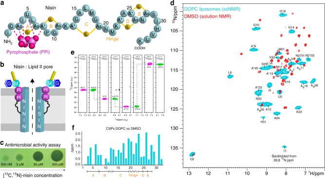
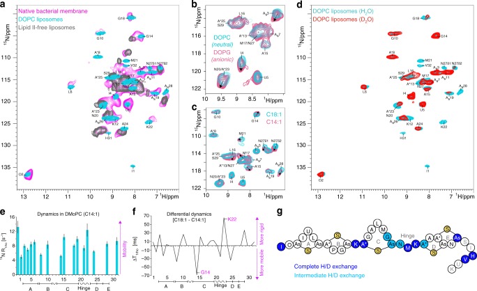
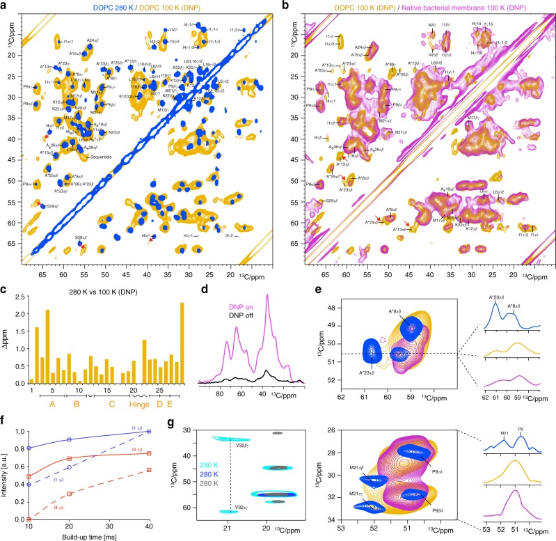
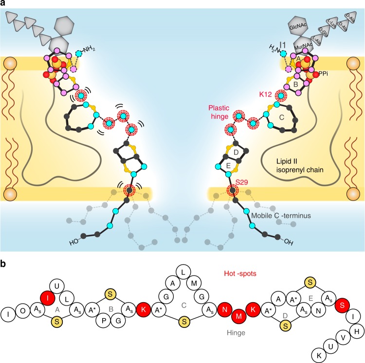
The alarming rise of antimicrobial resistance requires antibiotics with unexploited mechanisms. Ideal templates could be antibiotics that target the peptidoglycan precursor lipid II, known as the bacterial Achilles heel, at an irreplaceable pyrophosphate group. Such antibiotics would kill multidrug-resistant pathogens at nanomolecular concentrations without causing antimicrobial resistance. However, due to the challenge of studying small membrane-embedded drug-receptor complexes in native conditions, the structural correlates of the pharmaceutically relevant binding modes are unknown. Here, using advanced highly sensitive solid-state NMR setups, we present a high-resolution approach to study lipid II-binding antibiotics directly in cell membranes. On the example of nisin, the preeminent lantibiotic, we show that the native antibiotic-binding mode strongly differs from previously published structures, and we demonstrate that functional hotspots correspond to plastic drug domains that are critical for the cellular adaptability of nisin. Thereby, our approach provides a foundation for an improved understanding of powerful antibiotics.


Conflict of interest statement

The authors declare no competing interests.

Figures

Fig. 1
Fig. 1
ssNMR experiments of the lipid II–nisin complex in DOPC liposomes. a Illustration of the nisin Z peptide–antibiotic. The thio-ether rings are named A–E. Rings A–B are supposed to interact via five hydrogen bonds with lipid II. The non-canonical residues As, A*, O, and U are described in Supplementary Fig. 3. b Nisin and lipid II form a defined pore that spans the bacterial plasmamembrane. See Supplementary Fig. 3 for the lipid II structure. c A “spot-on-the-lawn” assay shows antimicrobial activity of [13C,15N]-labeled nisin. d Overlay of 1H-detected 2D 15N–1H spectra of the nisin–lipid II complex in DOPC (blue) and DMSO (red). We measured the ssNMR spectrum of nisin bound to lipid II in the pore state (2:1 stoichiometry) in DOPC at 950 MHz (1H-frequency) and 60 kHz MAS. The solution NMR spectrum in DMSO was previously published and shows a 1:1 nisin:lipid II complex. e Sequential assignments of lipid II-bound nisin in liposomes. Strip plots are shown from 3D CANH (magenta) and CAcoNH (green) ssNMR experiments. f Chemical shift perturbations (CSPs) comparing lipid II-bound nisin in DOPC and DMSO. CSPs were calculated according to CSP=Δ1H2+Δ15N6.512
Fig. 2
Fig. 2
The lipid II–nisin complex in cellular membranes. a Comparison of 1H-detected 2D 15N–1H spectra of lipid II-bound nisin in native M. flavus membranes (magenta) and in DOPC (cyan). The gray spectrum shows nisin non-specifically bound to DOPG:DOPC liposomes (7:3 ratio) in the absence of lipid II. b Overlay of 2D 15N–1H spectra of lipid II-bound nisin acquired in zwitterionic DOPC (cyan) and anionic DOPG:DOPC (7:3 ratio) (pink) liposomes. c Overlay of 2D 15N–1H spectra of lipid II-bound nisin acquired in C18:1 DOPC (cyan) and C14:1 DMoPC (pink) liposomes. d 1H/2H exchange: 2D 15N–1H spectra of lipid II-bound nisin acquired in DOPC in fully protonated (cyan) and deuterated (red) buffers. e 15N R1rho relaxation rates of lipid II-bound nisin acquired in DOPC. The error bars indicate the standard deviation of the fit. f Differential 15N T1rho times comparing dynamics in C18:1 DOPC and C14:1 DMoPC liposomes. g 1H/2H ssNMR exchange results. Dark and light blue beads represent residues that showed complete and intermediate exchange in deuterated buffers, respectively. Residues S29/I30 and U33/K34 could not be analyzed due to spectral overlap and fast dynamics, respectively
Fig. 3
Fig. 3
DNP-enhanced ssNMR on the lipid II-bound state of nisin in the pore. a 2D 13C–13C ssNMR spin diffusion spectra of the lipid II-bound state of nisin in the pore in DOPC acquired at 280 K sample temperature (blue) and at 100 K with DNP enhancement (orange). Red arrows highlight strong CSPs. b Overlay of DNP-ssNMR 2D 13C–13C spectra of nisin in the pore acquired in DOPC (orange) and in cellular M. flavus vesicles (magenta). Both spectra were acquired at identical conditions using 10.6 kHz MAS, 800 MHz and 40 ms 13C–13C mixing. c Combined (Cα + Cβ) CSPs comparing lipid II-bound nisin in DOPC at 280 K against 100 K temperature (DNP conditions). d 13C cross-polarization spectra of lipid II-bound nisin in M. flavus vesicles with (magenta) and without (black) DNP enhancement. e The hinge domain is conformationally flexible and broadens out at DNP conditions in DOPC (orange). This is even more pronounced in cellular membranes (magenta). Upper panel: (left) zoom into A*23 adjacent to the hinge; (right) slice through the A*23αβ signal. Lower panel: (left) zoom into M21; (right) projection along the indirect dimension (26–34 13C ppm). f 13C–13C PDSD spin diffusion buildup curves of the Cαβ (continuous lines) and Cαδ (dashed lines) cross-peaks for residues I1 (in blue) and I4 (red). g The mobile nisin C terminus gives faint signals in dipolar 2D 13C–13C spectra at 280 K (in gray and dark blue with 100 ms and 30 ms 13C–13C mixing, respectively), while stronger signals appear at 250 K (in cyan, 50 ms mixing)
Fig. 4
Fig. 4
The nisin:lipid II pore topology. a Membrane arrangement of the nisin:lipid II topology as seen by ssNMR. Plastic residues that are required to adapt to the bacterial target membrane are highlighted with red circles. Residues that showed 1H/2H exchange are colored in blue and align the pore lumen. The C terminus is dynamically disordered and resides at the water–membrane interface. The A–B rings (in magenta) interact with the lipid II PPi group. b Residues I4, K12, N20–K22, , , , , and S29 are pharmaceutical hotspots that enable to improve nisin’s activity upon mutation. These residues were all identified as important for nisin’s cellular adaptability in this study

References

    1. O’Neill, J. Antimicrobial Resistance: Tackling a Crisis for the Health and Wealth of Nations (Review on Antimicrobial Resistance, London, 2014).
    1. Breukink E, de Kruijff B. Lipid II as a target for antibiotics. Nat. Rev. Drug Discov. 2006;5:321–332. doi: 10.1038/nrd2004. - DOI - PubMed
    1. Schneider T, et al. Plectasin, a fungal defensin, targets the bacterial cell wall precursor Lipid II. Science. 2010;328:1168–1172. doi: 10.1126/science.1185723. - DOI - PubMed
    1. Cochrane SA, et al. Antimicrobial lipopeptide tridecaptin A1 selectively binds to Gram-negative lipid II. Proc. Natl Acad. Sci. USA. 2016;113:11561–11566. doi: 10.1073/pnas.1608623113. - DOI - PMC - PubMed
    1. Ling, L. L. et al. A new antibiotic kills pathogens without detectable resistance. Nature517, 455–459 (2015). - PMC - PubMed

Publication types