

MiR27a Promotes the Development of Macrophage-like Characteristics in 3T3-L1 Preadipocytes
- PMID: 30263011
- PMCID: PMC6158720
- DOI: 10.7150/ijbs.26274
MiR27a Promotes the Development of Macrophage-like Characteristics in 3T3-L1 Preadipocytes
Abstract
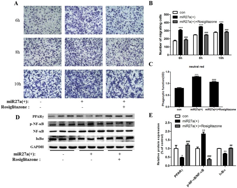
Recruitment and polarization of classically activated (M1) macrophages within adipose tissue contribute to chronic low-grade inflammation in obesity. Adipose tissue precursor cells exhibit the capacity to develop macrophage-like characteristics and adipocyte-derived miR27a is known to promote reprogramming of somatic cells. It was unknown whether exogenous addition of miR27a promote the development of macrophage-like characteristics of adipose precursor cells. We examined macrophage surface antigen, phagocytosis and migration ability in 3T3-L1 preadipocytes transfected with miR27a mimics. Transfection of 3T3-L1 preadipocytes with miR27a mimics increased phagocytosis and migration and increased the number of cells expressing the macrophage makers F4/80 and MHC compared to controls. M2 and CD206 macrophage markers were unaltered. In addition, transfection of 3T3-L1 preadipocytes with miR27a mimics reduced PPARγ expression, activated NF-κB and promoted secretion of the inflammatory cytokines MCP-1, TNF-α and IL-1β compared to controls. The level of anti-inflammatory factors Arg-1, IL-10, Ym1 and Fizz1 were unaltered. Secretion of miR27a was increased in conditioned medium prepared from palmitic acid-treated differentiated 3T3-L1 adipocytes compared to controls. Incubation of 3T3-L1 preadipocytes with this conditioned medium increased phagocytosis and migration compared to controls. Finally, conditioned medium prepared from differentiated 3T3-L1 adipocytes transfection with miR27a inhibitors reduced phagocytosis and migration in 3T3-L1 preadipocytes compared to controls. The data indicate that PPARγ agonists may reverse the activation of NF-κB pathway mediated by miR27a overexpression and reduce phagocytosis and migration of adipose precursor cells. In addition, miR27a may promote the development of macrophage-like characteristics in 3T3-L1 preadipocytes.
Keywords: 3T3 cells; inflammation; macrophage-like; miR27a; obesity; preadipocytes.
Conflict of interest statement
Competing Interests: The authors have declared that no competing interest exists.
Figures


Similar articles
-
α-Naphthoflavone modulates inflammatory response in adipocytes-macrophages interaction through NFκB signaling.Int J Clin Exp Pathol. 2014 Oct 15;7(11):7768-74. eCollection 2014. Int J Clin Exp Pathol. 2014. PMID: 25550814 Free PMC article.
-
The inhibitory effect of pterostilbene on inflammatory responses during the interaction of 3T3-L1 adipocytes and RAW 264.7 macrophages.J Agric Food Chem. 2013 Jan 23;61(3):602-10. doi: 10.1021/jf304487v. Epub 2013 Jan 11. J Agric Food Chem. 2013. PMID: 23268743
-
Role of the adipocyte-specific NF-κB activity in the regulation of IP-10 and T cell migration.Am J Physiol Endocrinol Metab. 2011 Feb;300(2):E304-11. doi: 10.1152/ajpendo.00143.2010. Epub 2010 Nov 9. Am J Physiol Endocrinol Metab. 2011. PMID: 21062959
-
Message Transmission Between Adipocyte and Macrophage in Obesity.Adv Exp Med Biol. 2024;1460:273-295. doi: 10.1007/978-3-031-63657-8_9. Adv Exp Med Biol. 2024. PMID: 39287855 Review.
-
Adipocyte-Macrophage Cross-Talk in Obesity.Adv Exp Med Biol. 2017;960:327-343. doi: 10.1007/978-3-319-48382-5_14. Adv Exp Med Biol. 2017. PMID: 28585206 Review.
Cited by
-
The role of RUNX1/NF-κB in regulating PVAT inflammation in aortic dissection.Sci Rep. 2024 Apr 30;14(1):9960. doi: 10.1038/s41598-024-60737-9. Sci Rep. 2024. PMID: 38693222 Free PMC article.
-
Blocking TRPV4 Ameliorates Osteoarthritis by Inhibiting M1 Macrophage Polarization via the ROS/NLRP3 Signaling Pathway.Antioxidants (Basel). 2022 Nov 23;11(12):2315. doi: 10.3390/antiox11122315. Antioxidants (Basel). 2022. PMID: 36552524 Free PMC article.
-
Long non-coding RNA (LncRNA) and epigenetic factors: their role in regulating the adipocytes in bovine.Front Genet. 2024 Oct 3;15:1405588. doi: 10.3389/fgene.2024.1405588. eCollection 2024. Front Genet. 2024. PMID: 39421300 Free PMC article. Review.
-
Exosome-guided direct reprogramming of tumor-associated macrophages from protumorigenic to antitumorigenic to fight cancer.Bioact Mater. 2022 Aug 5;25:527-540. doi: 10.1016/j.bioactmat.2022.07.021. eCollection 2023 Jul. Bioact Mater. 2022. PMID: 37056267 Free PMC article.
-
Alliin inhibits adipocyte differentiation by downregulating Akt expression: Implications for metabolic disease.Exp Ther Med. 2021 Jun;21(6):563. doi: 10.3892/etm.2021.9995. Epub 2021 Mar 26. Exp Ther Med. 2021. PMID: 33850535 Free PMC article.
References
-
- Francés DE, Motiño O, Agrá N. et al. Hepatic cyclooxygenase-2 expression protects against diet-induced steatosis, obesity and insulin resistance. Diabetes. 2015;64:1522–1531. - PubMed
-
- Curat CA, Miranville A, Sengenès C. et al. From Blood Monocytes to Adipose Tissue-Resident Macrophages: Induction of Diapedesis by Human Mature Adipocytes. Diabetes. 2004;53:1285–1292. - PubMed
-
- Katz S, Zsiros V, Dóczi N. et al. GM-CSF and GM-CSF receptor have regulatory role in transforming rat mesenteric mesothelial cells into macrophagelike cells. Inflamm Res. 2016;65:827–836. - PubMed
-
- Cousin B, Munoz O, Andre M. et al. A role for preadipocytes as macrophage-like cells. FASEB J. 1999;13:305–312. - PubMed
Publication types
MeSH terms
Substances
LinkOut - more resources
Full Text Sources
Other Literature Sources
Research Materials
Miscellaneous
