. 2018 Sep 24;6(5):e00432.
doi: 10.1002/prp2.432. eCollection 2018 Oct.

Real-time examination of cAMP activity at relaxin family peptide receptors using a BRET-based biosensor

Affiliations

Real-time examination of cAMP activity at relaxin family peptide receptors using a BRET-based biosensor

Adam L Valkovic et al. Pharmacol Res Perspect. .

Abstract

Relaxin family peptide (RXFPs) 1-4 receptors modulate the activity of cyclic adenosine monophosphate (cAMP) to produce a range of physiological functions. RXFP1 and RXFP2 increase cAMP via Gαs, whereas RXFP3 and RXFP4 inhibit cAMP via Gαi/o. RXFP1 also shows a delayed increase in cAMP downstream of Gαi3. In this study we have assessed whether the bioluminescence resonance energy transfer (BRET)-based biosensor CAMYEL (cAMP sensor using YFP-Epac-Rluc), which allows real-time measurement of cAMP activity in live cells, will aid in understanding ligand- and cell-specific RXFP signaling. CAMYEL detected concentration-dependent changes in cAMP activity at RXFP1-4 in recombinant cell lines, using a variety of ligands with potencies comparable to those seen in conventional cAMP assays. We used RXFP2 and RXFP3 antagonists to demonstrate that CAMYEL detects dynamic changes in cAMP by reversing cAMP activation or inhibition respectively, with real-time addition of antagonist after agonist stimulation. To demonstrate the utility of CAMYEL to detect cAMP activation in native cells expressing low levels of RXFP receptor, we cloned CAMYEL into a lentiviral vector and transduced THP-1 cells, which express low levels of RXFP1. THP-1 CAMYEL cells demonstrated robust cAMP activation in response to relaxin. However, the CAMYEL assay was unable to detect the Gαi3-mediated phase of RXFP1 cAMP activation in PTX-treated THP-1 cells or HEK293A cells with knockout of Gαs. Our data demonstrate that cytoplasmically-expressed CAMYEL efficiently detects real-time cAMP activation by Gαs or inhibition by Gαi/o but may not detect cAMP generated in specific intracellular compartments such as that generated by Gαi3 upon RXFP1 activation.

Keywords: CAMYEL; GPCR; RXFP; cAMP; relaxin; signalling.


Figures

Figure 1
Figure 1
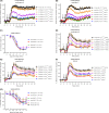
Ligand‐induced cAMP activity in HEKRXFP1 cells. HEK293T cells stably expressing RXFP1 were stimulated with vehicle, forskolin, or an RXFP1 agonist for 45 minutes. (A) Time‐course of forskolin‐induced cAMP activity. (B) Concentration‐response curve for forskolin at 45 minutes. Time‐course for (C) H2 relaxin‐ and (D) ML290‐induced cAMP activity. (E) Concentration‐response curves for H2 relaxin and ML290 at 5 and 45 minutes. Time of ligand or vehicle addition is represented by an arrow. Data represent mean ± SEM of three independent experiments
Figure 2
Figure 2
Ligand‐induced cAMP activity in HEKRXFP2 cells. HEK293T cells stably expressing RXFP2 were stimulated with ligands for 45 minutes. (A) Time‐course of forskolin‐induced cAMP activity. (B) Concentration‐response curve for forskolin at 45 minutes. Time‐course for (C) INSL3‐ and (D) H2 relaxin‐induced cAMP activity. (E) Concentration‐response curves for INSL3 and H2 relaxin at 5 and 45 minutes. (F) Blocking and reversibility of the CAMYEL signal was demonstrated by adding an RXFP2 antagonist (INSL3 B‐chain dimer) 10 minutes before or after agonist addition. Time of ligand or vehicle addition is represented by an arrow. Data represent mean ± SEM of three independent experiments
Figure 3
Figure 3
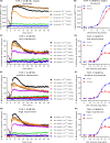
Ligand‐induced cAMP activity in cells expressing RXFP3. CHO‐K1 and HEK293T cells stably expressing RXFP3 (CHORXFP3 and HEKRXFP3) were stimulated with vehicle, forskolin, and an RXFP3 agonist for 45 minutes. CHORXFP3 cells were stimulated with (A) analogue 2 or (B) peptide 5, with addition of forskolin (10 μmol L−1) after 4 minutes. (C) Concentration‐response curves were generated for CHORXFP3 at 10 and 45 minutes. HEKRXFP3 cells were stimulated with (D) analogue 2, (E) peptide 5, or (C) R3/I5, with addition of forskolin (500 nmol L−1) after five minutes. (F) Concentration‐response curves were generated for HEKRXFP3 cells at 10 and 45 minutes. Time of agonist or vehicle (A) and forskolin or vehicle (F) addition are represented by arrows. Concentration‐response data is expressed as a percentage of forskolin for the indicated time point. Data represent mean ± SEM of three independent experiments
Figure 4
Figure 4
Blocking and reversing the CAMYEL signal in HEKRXFP3 cells. (A) Pre‐treatment of HEKRXFP3 cells with the Gαi/o inhibitor pertussis toxin (100 ng mL−1; 20 hours) abolished R3/I5‐induced inhibition of cAMP activity. (B) Treatment with the RXFP3 antagonist R3 B1‐22R after stimulation with R3/I5 (30 minutes) and forskolin showed reversal of the R3/I5 response in real time. Time of ligand or vehicle addition is represented by an arrow. Data represent the mean ± SEM of three independent experiments
Figure 5
Figure 5
Ligand‐induced cAMP activity in CHORXFP4 cells. CHO‐K1 cells stably expressing RXFP4 (CHORXFP4) were stimulated with vehicle, forskolin, and an RXFP4 agonist for 45 minutes, with addition of forskolin (10 μmol L−1) 4 minutes after agonist addition. Time‐courses were generated for (A) INSL5 and (B) analogue 13. (C) Concentration‐response curves were generated for both agonists at 10 and 45 minutes. Time of agonist or vehicle (A) and forskolin or vehicle (F) addition are represented by arrows. Concentration‐response data is expressed as a percentage of forskolin for the indicated time point. Data represent mean ± SEM of three independent experiments
Figure 6
Figure 6
Representation of fluorescence‐activated cell sorting (FACS) of transduced THP‐1 CAMYEL cells. THP‐1 cells were transduced by spinoculation with CAMYEL lentivirus and were sorted to remove untransduced cells. Single, live cells were isolated, and sorted by expression of YFP into low, medium, and high levels of CAMYEL expression, based on YFP fluorescence. Cells with YFP expression that was no greater than control THP‐1 cells were removed
Figure 7
Figure 7
Ligand‐induced cAMP activity in THP‐1 CAMYEL cells. THP‐1 cells that endogenously express RXFP1 and stably express high, medium, or low levels of CAMYEL (THP‐1 CAMYEL) were stimulated with vehicle, forskolin, or H2 relaxin for 45 minutes. (A) Time‐course for forskolin activity in high expression cells. (B) Concentration‐response curves for forskolin at 5 and 45 minutes. (C, E, G) Time‐courses for H2 relaxin‐induced cAMP activation in the low‐, medium‐, and high‐expressing cells. (D, F, H) Concentration‐response curves for H2 relaxin at 5 and 45 minutes in the low‐, medium‐, and high‐expressing cells. Time of ligand or vehicle addition is represented by an arrow. Data represent mean ± SEM of three independent experiments
Figure 8
Figure 8
Effect of pertussis toxin on cAMP activity in THP‐1 CAMYEL cells. THP‐1 CAMYEL (high expression) were either (A) untreated or (B) treated with pertussis toxin (100 ng mL−1; 16‐18 hours), and stimulated with vehicle, forskolin, or H2 relaxin for 45 minutes. Treated and untreated cells were compared at (C) 5 and (D) 45 minutes. Time of ligand or vehicle addition is expressed by an arrow. Data in (C) and (D) are expressed as a percentage of the forskolin response at the same time point. Data represent the mean ± SEM of four independent experiments
Figure 9
Figure 9
cAMP activity in HEK293A parental and Gαs knockout (ΔGαs) cells. (A) HEK293A parental cells, (B) HEK293A ΔGαs cells, and (C) HEK293A ΔGαs cells with Gαs transfected in, were transfected with CAMYEL and stimulated with vehicle or forskolin for 45 minutes. (D) Concentration‐response curves were generated for forskolin at 5 and 45 minutes. (E) HEK293A parental and (F) HEK293A ΔGαs cells were co‐transfected with CAMYEL and RXFP1, and were stimulated with vehicle, forskolin, isoprenaline, or H2 relaxin for 45 minutes. (G) HEK293A ΔGαs cells were primed with forskolin (33 μmol L−1) to compensate for the loss of Gαs, before adding forskolin, isoprenaline, or H2 relaxin. Time of ligand or vehicle addition is represented by an arrow. Data represent mean ± SEM of three independent experiments

Similar articles

Cited by

References

    1. Rajagopal S, Rajagopal K, Lefkowitz RJ. Teaching old receptors new tricks: biasing seven‐transmembrane receptors. Nat Rev Drug Discov. 2010;9:373‐386. - PMC - PubMed
    1. Smith JS, Lefkowitz RJ, Rajagopal S. Biased signalling: from simple switches to allosteric microprocessors. Nat Rev Drug Discov. 2018;17:243‐260. - PMC - PubMed
    1. Bathgate RA, Kocan M, Scott DJ, et al. The relaxin receptor as a therapeutic target – perspectives from evolution and drug targeting. Pharmacol Ther. 2018;187:114‐132. - PubMed
    1. Bathgate RAD, Halls ML, van der Westhuizen ET, Callander GE, Kocan M, Summers RJ. Relaxin family peptides and their receptors. Physiol Rev. 2013;93:405‐480. - PubMed
    1. Bathgate RA, Ivell R, Sanborn BM, Sherwood OD, Summers RJ. International Union of Pharmacology LVII: recommendations for the nomenclature of receptors for relaxin family peptides. Pharmacol Rev. 2006;58:7‐31. - PubMed

Publication types

LinkOut - more resources