

MRI of lobar holoprosencephaly in a cat with hypodipsic hypernatraemia
- PMID: 30263144
- PMCID: PMC6149025
- DOI: 10.1177/2055116918801602
MRI of lobar holoprosencephaly in a cat with hypodipsic hypernatraemia
Abstract
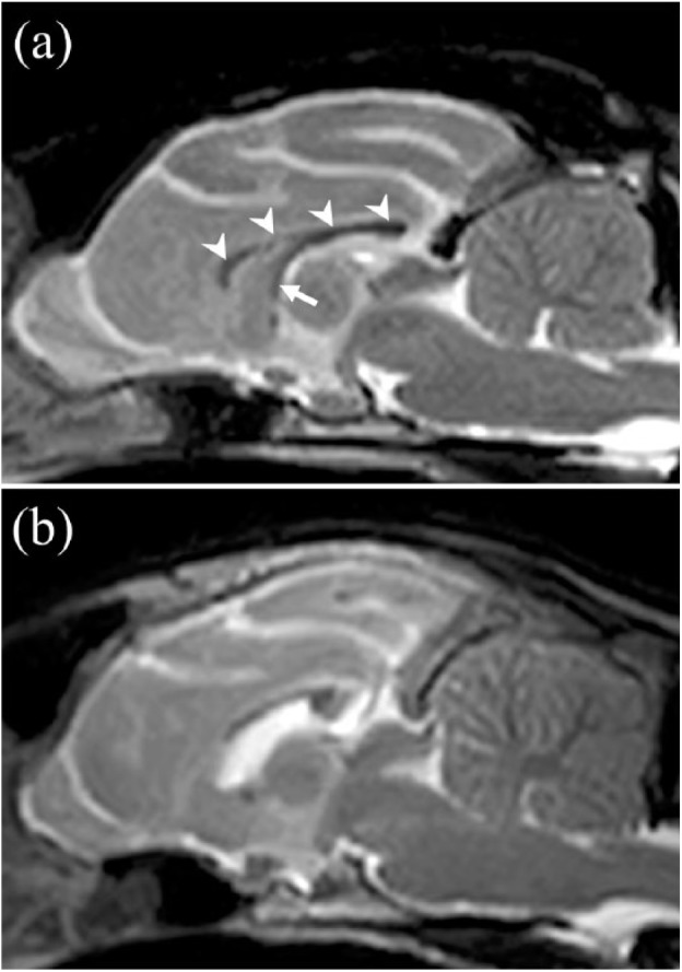
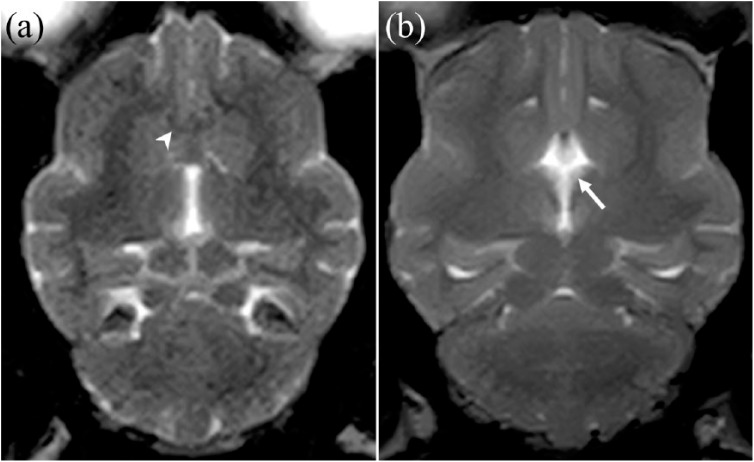
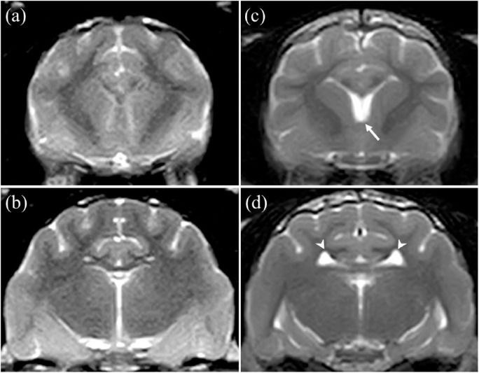
Case summary: A 2-year-old neutered female domestic shorthair cat presented with a history of hypodipsia, recurrent hypernatraemia, pelvic limb ataxia and tremor. The serum arginine vasopressin level was low for the serum osmolality. MRI of the brain revealed a failure of separation of the cerebrum, which manifested as absence of the rostral part of the corpus callosum, fornix and septum pellucidum, thus resulting in a single fused ventricle. The diagnosis was lobar holoprosencephaly with hypodipsic hypernatraemia.
Relevance and novel information: To our knowledge, this is the first description of the MRI characteristics of lobar holoprosencephaly in a cat. This report suggests that MRI examination should be considered for precise diagnosis of hypodipsic hypernatraemia in young cats.
Keywords: Brain malformation; holoprosencephaly; hypernatraemia; hypodipsia; magnetic resonance imaging.
Conflict of interest statement
Conflict of interest: The authors declared no potential conflicts of interest with respect to the research, authorship, and/or publication of this article.
Figures
Similar articles
-
Successful treatment of hypodipsic/adipsic hypernatremia in a cat with lobar holoprosencephaly using oral desmopressin.JFMS Open Rep. 2022 Mar 23;8(1):20551169221082542. doi: 10.1177/20551169221082542. eCollection 2022 Jan-Jun. JFMS Open Rep. 2022. PMID: 35342639 Free PMC article.
-
Hypodipsia-hypernatremia syndrome associated with holoprosencephaly in a child: a case report.Turk J Pediatr. 2002 Jul-Sep;44(3):263-6. Turk J Pediatr. 2002. PMID: 12405444
-
Semilobar Holoprosencephaly with Neurogenic Hypernatraemia: Two new cases.Sultan Qaboos Univ Med J. 2013 Aug;13(3):E463-6. Epub 2013 Jun 25. Sultan Qaboos Univ Med J. 2013. PMID: 23984038 Free PMC article.
-
Holoprosencephaly and septo-optic dysplasia.Neuroimaging Clin N Am. 1994 May;4(2):263-81. Neuroimaging Clin N Am. 1994. PMID: 8081628 Review.
-
[Cerebral malformation in the newborn: holoprosencephaly and agenesis of the corpus callosum].Rev Neurol. 2003 Jan 16-31;36(2):179-84. Rev Neurol. 2003. PMID: 12589607 Review. Spanish.
Cited by
-
Successful treatment of hypodipsic/adipsic hypernatremia in a cat with lobar holoprosencephaly using oral desmopressin.JFMS Open Rep. 2022 Mar 23;8(1):20551169221082542. doi: 10.1177/20551169221082542. eCollection 2022 Jan-Jun. JFMS Open Rep. 2022. PMID: 35342639 Free PMC article.
-
Case Report: Hypodipsic hypernatremia secondary to hydrocephalus in a dog.Front Vet Sci. 2025 May 16;12:1579965. doi: 10.3389/fvets.2025.1579965. eCollection 2025. Front Vet Sci. 2025. PMID: 40454171 Free PMC article.
-
Congenital pituitary cyst resulting in adipsic central diabetes insipidus and secondary hypernatremia in a cat.JFMS Open Rep. 2021 Feb 27;7(1):2055116921990294. doi: 10.1177/2055116921990294. eCollection 2021 Jan-Jun. JFMS Open Rep. 2021. PMID: 33738109 Free PMC article.
-
Hypodipsic hypernatremia after long-standing polydipsia in a cat with suspect neonatal head trauma.Can Vet J. 2023 Nov;64(11):1021-1027. Can Vet J. 2023. PMID: 37915774 Free PMC article.
-
A Deletion in GDF7 is Associated with a Heritable Forebrain Commissural Malformation Concurrent with Ventriculomegaly and Interhemispheric Cysts in Cats.Genes (Basel). 2020 Jun 19;11(6):672. doi: 10.3390/genes11060672. Genes (Basel). 2020. PMID: 32575532 Free PMC article.
References
-
- Dugger DT, Mellema MS, Hopper K, et al. Comparative accuracy of several published formulae for the estimation of serum osmolality in cats. J Small Anim Pract 2013; 54: 184–189. - PubMed
-
- Brown BA, Peterson ME, Robertson GL. Evaluation of the plasma vasopressin, plasma sodium, and urine osmolality response to water restriction in normal cats and a cat with diabetes insipidus. J Vet Intern Med 1993; 7: 113.
-
- Hahn JS, Barnes PD. Neuroimaging advances in holoprosencephaly: refining the spectrum of the midline malformation. Am J Med Genet C Semin Med Genet 2010; 154: 120–132. - PubMed
-
- Scott FW, LaHunta A, Schultz RD, et al. Teratogenesis in cats associated with griseofulvin therapy. Teratology 1975; 11: 79–86. - PubMed
Publication types
LinkOut - more resources
Full Text Sources
Other Literature Sources
Miscellaneous