

The suppressive effect of Gelidium amansi- EtOH extracts on the adipogenesis with MAPK signals in adipocytes with or without macrophages
- PMID: 30263710
- PMCID: PMC6049707
- DOI: 10.1007/s10068-017-0230-z
The suppressive effect of Gelidium amansi- EtOH extracts on the adipogenesis with MAPK signals in adipocytes with or without macrophages
Abstract
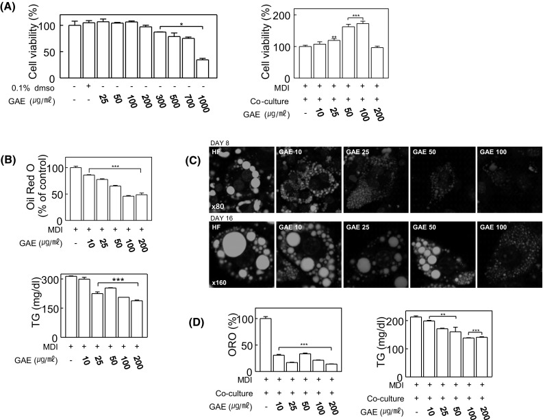
To elucidate the anti-inflammatory and anti-adipogenetic effects of Gelidium amansii (GA) ethanol extracts and their mechanisms, we performed two culture systems, adipocytes cultured with or without macrophages. Purified GA-3 fraction (GAE) contains high flavonoids and phenolics, reduced the mRNA levels of PPARγ and C/EBPα with GLUT4 expression in adipocyte with or without macrophages. GAE also increased the protein expression of HSL and ATGL enzymes, lipolysis biomarkers in fat cells. In co-culture system, GAE suppressed not only the transcription factors for adipogenesis, but also the production of pro-inflammatory cytokines, TNF-α. Compared to MAPK pathways such as JNK and p-38, the phosphorylation of both ERK1/2 (Thr202/Tyr204) was strongly suppressed by GAE with dose-dependent manner in both culture system. Otherwise, an increased JNK expression caused by GAE treatments blocked an insulin-induced GLUT4 translocation in adipocytes culture. In conclusion, GAE depressed the expression of adipogenetic genes, corresponding to a reduction in fat accumulation while preadipocytes developed into adipocytes with the modulation of MAPK pathways and inflammatory cytokines.
Keywords: Adipocytes; ERK1/2; Gelidium amansii; MAPK; Macrophages; TNF-α.
Conflict of interest statement
Compliance with ethical standardsThe authors declare no conflict of interest.
Figures


Similar articles
-
Popular edible seaweed, Gelidium amansii prevents against diet-induced obesity.Food Chem Toxicol. 2016 Apr;90:181-7. doi: 10.1016/j.fct.2016.02.014. Epub 2016 Feb 18. Food Chem Toxicol. 2016. PMID: 26911551
-
Effects of orexin A on GLUT4 expression and lipid content via MAPK signaling in 3T3-L1 adipocytes.J Steroid Biochem Mol Biol. 2013 Nov;138:376-83. doi: 10.1016/j.jsbmb.2013.07.005. Epub 2013 Jul 29. J Steroid Biochem Mol Biol. 2013. PMID: 23907013
-
Gelidium amansii ethanol extract suppresses fat accumulation by down-regulating adipogenic transcription factors in ob/ob mice model.Food Sci Biotechnol. 2017 Feb 28;26(1):207-212. doi: 10.1007/s10068-017-0028-z. eCollection 2017. Food Sci Biotechnol. 2017. PMID: 30263530 Free PMC article.
-
Suppression of adipocyte hypertrophy by polymethoxyflavonoids isolated from Kaempferia parviflora.Phytomedicine. 2014 May 15;21(6):800-6. doi: 10.1016/j.phymed.2014.01.014. Epub 2014 Mar 11. Phytomedicine. 2014. PMID: 24629599
-
Extract from Edible Red Seaweed (Gelidium amansii) Inhibits Lipid Accumulation and ROS Production during Differentiation in 3T3-L1 Cells.Prev Nutr Food Sci. 2012 Jun;17(2):129-35. doi: 10.3746/pnf.2012.17.2.129. Prev Nutr Food Sci. 2012. PMID: 24471074 Free PMC article.
Cited by
-
The Antiproliferative and Apoptosis-Inducing Effects of the Red Macroalgae Gelidium latifolium Extract against Melanoma Cells.Molecules. 2021 Oct 30;26(21):6568. doi: 10.3390/molecules26216568. Molecules. 2021. PMID: 34770978 Free PMC article.
-
Sulfated Galactans from Agarophytes: Review of Extraction Methods, Structural Features, and Biological Activities.Biomolecules. 2023 Dec 5;13(12):1745. doi: 10.3390/biom13121745. Biomolecules. 2023. PMID: 38136616 Free PMC article. Review.
-
Anti-inflammatory effects of Agar free-Gelidium amansii (GA) extracts in high-fat diet-induced obese mice.Nutr Res Pract. 2018 Dec;12(6):479-485. doi: 10.4162/nrp.2018.12.6.479. Epub 2018 Oct 12. Nutr Res Pract. 2018. PMID: 30515275 Free PMC article.
-
Effect of (Poly)phenols on Lipid and Glucose Metabolisms in 3T3-L1 Adipocytes: an Integrated Analysis of Mechanistic Approaches.Curr Obes Rep. 2025 Aug 6;14(1):64. doi: 10.1007/s13679-025-00656-6. Curr Obes Rep. 2025. PMID: 40767889 Free PMC article. Review.
References
LinkOut - more resources
Full Text Sources
Other Literature Sources
Research Materials
Miscellaneous