. 2017 Dec 12;27(1):147-157.
doi: 10.1007/s10068-017-0276-y. eCollection 2018 Feb.

The anti-photoaging and moisturizing effects of Bouea macrophylla extract in UVB-irradiated hairless mice

Affiliations

The anti-photoaging and moisturizing effects of Bouea macrophylla extract in UVB-irradiated hairless mice

Yongin Cheong et al. Food Sci Biotechnol. .

Abstract

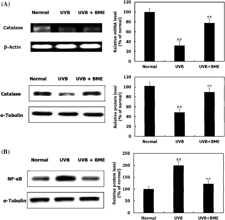
Ultraviolet (UV) light, a main cause of photoaging, leads to collapse of skin structure, resulting in wrinkle formation and dehydration. The present study assessed the anti-photoaging and moisturizing effects of Bouea macrophylla extract (BRE). UVB-irradiated hairless mice were orally administered with BME (300 mg/kg/day) for 8 weeks. BME ameliorated wrinkle formation, skin thickening, and inelasticity. BME upregulated COL1A1, COL3A1, COL4A1, and COL7A1 mRNA levels through activation of the transforming growth factor-β (TGF-β)/Smad pathway, thereby recovering the content of collagen reduced by UVB. Further, BME suppressed UVB-induced matrix metalloproteinase (MMP)-3 and MMP-13 expression and inhibited MMP-2 and MMP-9 activity by mediating the mitogen-activated protein kinases (MAPKs)/activator protein-1 (AP-1). BME improved moisture content by stimulating the expression of cornified envelope proteins and filaggrin-processing enzymes. Overall, the results show that BME prevents photoaging and promotes moisturization in UVB-irradiated hairless mice, suggesting its potential as a nutraceutical candidate for anti-photoaging and moisturizing effects.

Keywords: Anti-photoaging; Bouea macrophylla; Collagen; Matrix metalloproteinase; Moisturizing effect.


Conflict of interest statement

Compliance with ethical standardsThe authors declare no conflict of interest.

Figures

Fig. 1
Fig. 1
Effect of BME on UVB-induced photoaging symptoms. (A) Replica photographs indicating wrinkle formation and wrinkle parameters were determined by skin replica analysis. (B) Measurement of skinfold thickness and hematoxylin and eosin staining of dorsal skin tissue section. (C) Gross elasticity (Ua/Uf) of skin and Verhoeff’s staining of elastic fiber in dermis. Data are represented as the mean ± SD (n = 5). ## p < 0.01 (Normal group vs. UVB-irradiated group); **p < 0.01 (UVB-irradiated group vs. BME-treated group)
Fig. 2
Fig. 2
Effects of BME on catalase and NF-κB expression. (A) RT-PCR and western blot analysis indicating the expression of catalase at mRNA and protein levels. (B) Western blot analysis indicating the expression of NF-κB at protein level. Data are represented as the mean ± SD (n = 5). ## p < 0.01 (Normal group vs. UVB-irradiated group); **p < 0.01 (UVB-irradiated group vs. BME-treated group)
Fig. 3
Fig. 3
Effects of BME on collagen content and synthesis. (A) RT-PCR analysis indicating the expression of COL1A1, COL3A1, COL4A1, and COL7A1 mRNA. (B) Amount of hydroxyproline and Masson’s trichrome stain of collagen fibers. (C) Western blot analysis indicating the expression of TGF-β/Smad signaling pathway at protein level. Data are represented as the mean ± SD (n = 5). ## p < 0.01 (Normal group vs. UVB-irradiated group); **p < 0.01 (UVB-irradiated group vs. BME-treated group)
Fig. 4
Fig. 4
Effects of BME on UVB-induced MMPs activity and expression. (A) Gelatin zymography analysis indicating the activity of MMP-2 and MMP-9. (B) RT-PCR analysis indicating the mRNA expression levels of MMP-3 and MMP-13. (C) Western blot analysis indicating the protein expression of MMP-3 and MMP-13. Data are represented as the mean ± SD (n = 5). ## p < 0.01 (Normal group vs. UVB-irradiated group); **p < 0.01 (UVB-irradiated group vs. BME-treated group)
Fig. 5
Fig. 5
Effect of BME on UVB-induced MAPKs/AP-1 complex signaling pathway. Western blot analysis indicating the expression of (A) MAPKs and (B) AP-1 complex at protein levels. Data are represented as the mean ± SD (n = 5). ## p < 0.01 (Normal group vs. UVB-irradiated group); **p < 0.01 (UVB-irradiated group vs. BME-treated group)
Fig. 6
Fig. 6
Effects of BME on CE formation and filaggrin processing. (A) The mRNA and protein expression of loricrin, involucrin, and transglutaminase. (B) TEWL as a marker of skin dehydration in UVB-irradiated dorsal skin. (C) The mRNA and protein expression of filaggrin and caspase-14. (D) The protein levels of matriptase and prostasin. (E) Graph indicating skin hydration in UVB-irradiated dorsal skin. Data are represented as the mean ± SD (n = 5). ## p < 0.01 (Normal group vs. UVB-irradiated group); *p < 0.05, **p < 0.01 (UVB-irradiated group vs. BME-treated group)

References

    1. El-Domyati M, Attia S, Saleh F, Brown D, Birk DE, Gasparro F, Ahmad H, Uitto J. Intrinsic aging vs. photoaging: a comparative histopathological, immunohistochemical, and ultrastructural study of skin. Experi. Dermatol. 11: 398–405 (2002). - PubMed
    1. Kuhn A, Zahn S, Patsinakidis N, Landmann A, Graef M, Sauerland C, Surber C, Wenzel J. Resistance to water and abrasion of a broad-spectrum sunscreen: a prospective, open-label study. Experi. Dermatol. 2016;25:151–152. doi: 10.1111/exd.12869. - DOI - PubMed
    1. Bickers DR, Athar M. Oxidative stress in the pathogenesis of skin disease. J. Investig. Dermatol. 2006;126:2565–2575. doi: 10.1038/sj.jid.5700340. - DOI - PubMed
    1. Passeron T, Ortonne J. Skin ageing and its prevention. Press. Medica. 2003;32:1474–1482. - PubMed
    1. Heng MC. Signaling pathways targeted by curcumin in acute and chronic injury: burns and photo-damaged skin. Int. J. Dermatol. 2013;52:531–543. doi: 10.1111/j.1365-4632.2012.05703.x. - DOI - PubMed

LinkOut - more resources