

A case report of intracholecystic papillary neoplasm of the gallbladder resembling a submucosal tumor
- PMID: 30264362
- PMCID: PMC6160379
- DOI: 10.1186/s40792-018-0524-2
A case report of intracholecystic papillary neoplasm of the gallbladder resembling a submucosal tumor
Abstract
Background: Intracholecystic papillary neoplasm (ICPN) is defined as papillary tumors detected macroscopically in the gallbladder. We report a case of ICPN which exhibited the atypical form like a submucosal tumor.
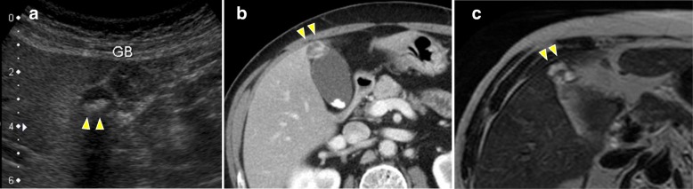
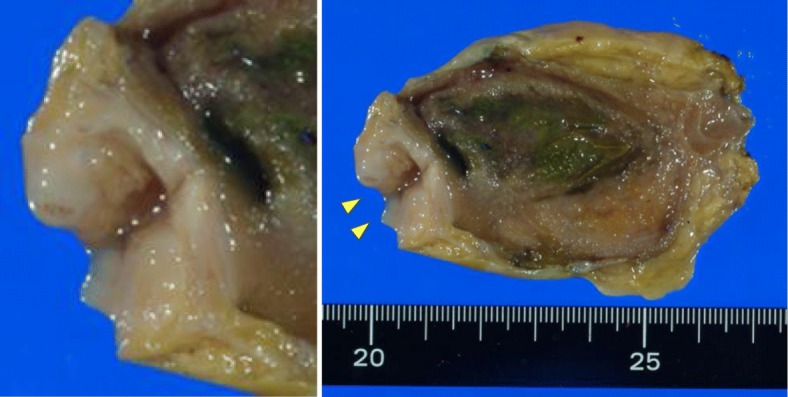
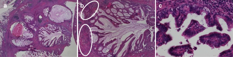
Case presentation: A 70-year-old man was admitted to our hospital because of hepatic disorder. Computed tomography and magnetic resonance imaging showed irregular thickening of the wall within the gallbladder fundus. Because the lesion might have been malignant, we performed laparoscopic cholecystectomy and liver bed resection. Macroscopic findings showed the mucosal surface of the tumor was smooth, and its form was similar to that of a submucosal tumor. Histopathological examination revealed papillary tumors within the mass with low-grade dysplasia; therefore, we diagnosed ICPN.
Conclusion: In the present case, ICPN was resembling a submucosal tumor macroscopically because the tumors arose into the Rokitansky-Aschoff sinus and the adenomyomatous hyperplasia was merged with the ICPN. It is necessary to consider the possibility of tumor lesions within adenomyomatous hyperplasia.
Keywords: Adenomyomatous hyperplasia; Intracholecystic papillary neoplasm; Laparoscopic cholecystectomy.
Conflict of interest statement
Ethics approval and consent to participate
Not applicable.
Consent for publication
Written informed consent was obtained from the patient for the publication of this case report and the accompanying images.
Competing interests
The authors declare that they have no competing interests.
Publisher’s Note
Springer Nature remains neutral with regard to jurisdictional claims in published maps and institutional affiliations.
Figures

References
-
- Albores-Saavedra J, Adsay NV, Crawford JM, Klimstra DS, Kloppel G, Sripa B, Tsui WMS, Paradis V. Carcinoma of the gallbladder and extrahepatic ducts. In: Bosman FT, Carneiro F, Hruban RH, Theise ND, editors. WHO classification of tumours of the digestive system. 4. Lyon: International Agency for Research on Cancer; 2010. pp. 266–273.
LinkOut - more resources
Full Text Sources
Other Literature Sources
