Review
. 2019 Dec;25(6):566-582.
doi: 10.1177/1073858418803594. Epub 2018 Sep 28.

Functional and Neuroanatomical Bases of Developmental Stuttering: Current Insights

Affiliations
Review

Functional and Neuroanatomical Bases of Developmental Stuttering: Current Insights

Soo-Eun Chang et al. Neuroscientist. 2019 Dec.

Abstract

Affecting 5% of all preschool-aged children and 1% of the general population, developmental stuttering-also called childhood-onset fluency disorder-is a complex, multifactorial neurodevelopmental disorder characterized by frequent disruption of the fluent flow of speech. Over the past two decades, neuroimaging studies of both children and adults who stutter have begun to provide significant insights into the neurobiological bases of stuttering. This review highlights convergent findings from this body of literature with a focus on functional and structural neuroimaging results that are supported by theoretically driven neurocomputational models of speech production. Updated views on possible mechanisms of stuttering onset and persistence, and perspectives on promising areas for future research into the mechanisms of stuttering, are discussed.

Keywords: DTI; MRI; neurodevelopmental disorder; speech; stuttering.


Figures
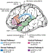
Figure 1.
Figure 1.
Speech-language cortical areas and the dorsal and ventral pathways that interconnect them. Multiple studies examining stuttering speakers have reported decreased white matter integrity in the left dorsal pathways, and morphological changes affecting the motor and auditory cortical areas that are interconnected by these tracts. AF/SLF: arcuate fasciculus/superior longitudinal fasciculus; BA: Brodmann area; EFCS: extreme fiber capsule system; FOP: frontal operculum; UF: uncinate fasciculus. Figure reprinted and modified with permission from The American Physiological Society and Copyright Clearance Center. The Brain Basis of Language Processing: From Structure to Function. Friederici AD. Physiological Reviews. 2011.
Figure 2.
Figure 2.
Convergent findings from past diffusion tensor imaging (DTI) studies of stuttering focus on deficits in sections along the left superior longitudinal fasciculus. Reprinted and adapted by permission from Springer Customer Service Centre GmbH: Springer Nature. The Neurobiological Grounding of Persistent Stuttering: from Structure to Function. Neef NE, Anwander A, Friederici AD. Current Neurology and Neuroscience Reports. 2015.
Figure 3.
Figure 3.
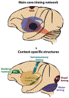
Major neural structures supporting estimation of time. The core structures include the basal ganglia and supplementary motor area (SMA). These structures interface with a distributed network of areas in a context specific manner. Reprinted and modified with permission from Annual Reviews, Inc: Annual Review of Neuroscience. Neural Basis of the Perception and Estimation of Time. Merchant H, Harrington DL, Meck WH. 2013.
Figure 4.
Figure 4.
While age-related increases in white matter integrity was observed in areas (left temporoparietal junction, posterior STG) along the left arcuate fasciculus in normally fluent children as well as children who recover from stuttering, the developmental trajectories in the same regions lacked age-related increases in children with persistent stuttering. Arc-fp: arcuate fasciculus-frontoparietal; arc-t: arcuate fasciculus-temporal; ilf: inferior longitudinal fasciculus; Slf: superior longitudinal fasciculus. Figure reprinted and modified with permission from John Wiley and Sons and Copyright Clearance Center. White matter developmental trajectories associated with persistence and recovery of childhood stuttering. Chow HM, Chang S-E. Human Brain Mapping, 2017.
Figure 5.
Figure 5.
Morphometric differences in speech motor control regions differentiated children with persistent stuttering from those who recover. A compensatory mechanism involving left medial premotor cortex may contribute to recovery. From “Anomalous morphology in left hemisphere motor and premotor cortex of children who stutter,” by Garnett et al., 2018, Brain, in press, Copyright 2018 by the Oxford University Press. Reprinted with permission.
Figure 6.
Figure 6.
A plausible neural framework relevant to stuttering risk, persistence, and recovery. Neural structures critical for sensorimotor integration for speech planning and production interface with subcortical structures that provide temporal structure and enable internal timing of speech sound production. Colors and thickness of lines are hypothetical, based on previous reported findings in the literature. Modified based on a subcortical-cortical model proposed by Kotz and colleagues, this model provides a useful framework that incorporates most convergent empirical findings to date on neural deficits linked to stuttering. Elucidating the functional and structural connectivity among component areas and causal relationships represented here could lead to novel insights into possible neural mechanisms linked to stuttering. BG: basal ganglia; caud: caudate; CE: cerebellum; IFG: inferior frontal gyrus; M1: primary motor cortex; PMC: premotor cortex; put: putamen; PWS: people who stutter; SMA: supplementary motor area; SMG/IPL: supramarginal gyrus/inferior parietal lobe; STG: superior temporal gyrus; vPMC: ventral premotor cortex.
Figure 7.
Figure 7.
Relationship between gene expression of two stuttering-linked genes (GNPTG and NAGPA) and absolute regional gray matter volume differences observed in persistent children who stutter. Dots represent brain regions in the left hemisphere defined by the AAL atlas. The brain regions with relatively high expression of GNPTG and NAGPA and between-group differences in GMV were primarily in the sensorimotor, parietal, the cingulate cortex, and the middle frontal gyrus. The Spearman’s rank correlation coefficients for gene expression-brain volume group difference were ρ = 0.57 for GNPTG, and ρ = 0.42 for NAGPA. Chow et al., 2018.

References

    1. Adams MR, Ramig P. 1980. Vocal characteristics of normal speakers and stutterers during choral reading. J Speech Hear Res 23:457–469. - PubMed
    1. Alm PA. 2004. Stuttering and the basal ganglia circuits: a critical review of possible relations. Journal of Communication Disorders 37:325–370. - PubMed
    1. Andrews CC, O’Brian SS, Harrison EE, Onslow MM, Packman AA, Menzies RR. 2012. Syllable-timed speech treatment for school-age children who stutter: a phase I trial. Language, Speech, and Hearing Services in Schools 43:359–369. - PubMed
    1. Andrews G, Harris M. 1964. The syndrome of stuttering. Spastics Society Medical Education:191.
    1. Bartolo R, Merchant H. 2015. β oscillations are linked to the initiation of sensory-cued movement sequences and the internal guidance of regular tapping in the monkey. J Neurosci [Internet] 35:4635–4640. Available from: http://www.ncbi.nlm.nih.gov/pubmed/?term=β+oscillations+are+linked+to+th.... - PMC - PubMed

Publication types