

Review
doi: 10.4103/ijmr.IJMR_1793_17.
Oral health consequences of smokeless tobacco use
Affiliations
- PMID: 30264752
- PMCID: PMC6172921
- DOI: 10.4103/ijmr.IJMR_1793_17
Item in Clipboard
Review
Oral health consequences of smokeless tobacco use
Indian J Med Res.
2018 Jul.
Abstract
Smokeless tobacco (SLT) use has many oral effects including oral cancer, leukoplakia and erythroplakia, oral submucous fibrosis (if mixed with areca nut), loss of periodontal support (recession) and staining of teeth and composite restorations. This review was aimed to provide information to identify oral lesions that occur due to the use of smokeless tobacco so that effective interventions can be undertaken to reduce morbidity and mortality from the use of SLT.
Keywords: Chewing tobacco - leukoplakia - oral cancer - smokeless tobacco - submucous fibrosis.
Conflict of interest statement
None
Figures
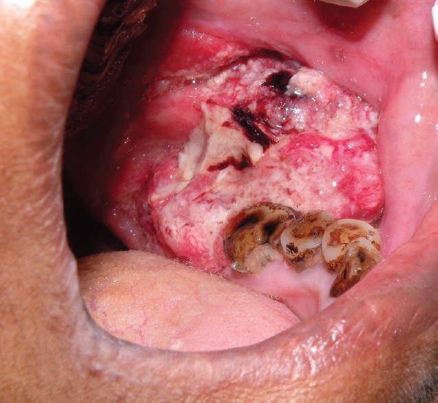
Oral squamous cell carcinoma in a patient addicted to gutka. Tumour arising from retromolar region has spread to the buccal sulcus. The figure demonstrates features of an exophytic growth with ulceration and necrosis.
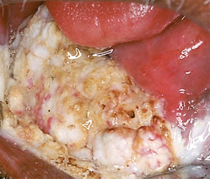
Verrucous carcinoma in oral cavity of a patient in a tobacco chewer. This is an advanced tumour with a typical verrucoid appearance.
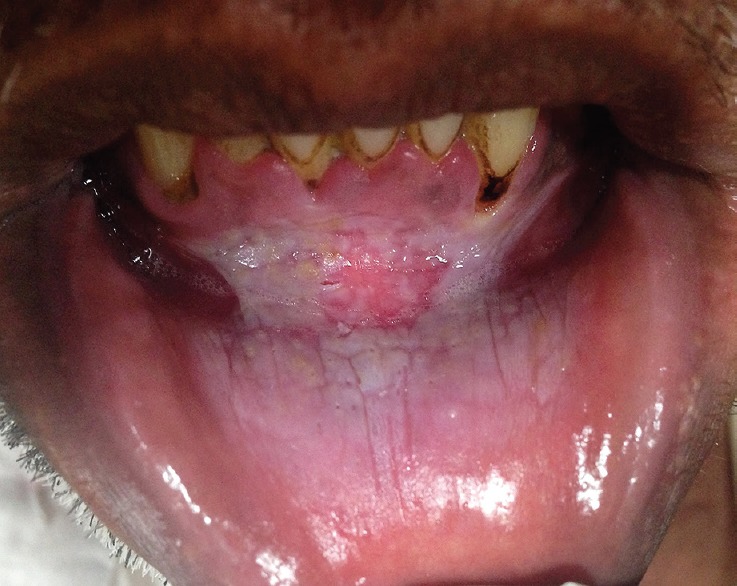
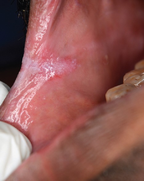
Lower labial sulcus of a patient showing tobacco pouch keratosis in places where the quid has been retained for long periods.
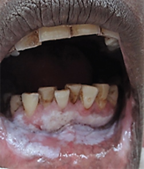
Oral homogeneous leukoplakia affecting the commisure in a tobacco chewer.
Erythroleukoplakia with candidal infection. This is mixed red and white lesion and has a worse prognosis than homogeneous leukoplakia.
Proliferative verrucous leukoplakia. Thick white plaques are noted affecting several sites, gingiva, alveolar mucosa, the sulcus and spreading to lower lip.
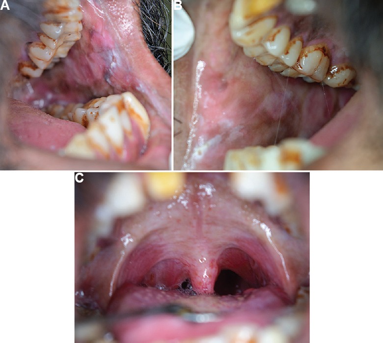
Oral submucous fibrosis, (A) left buccal mucosa, (B) right buccal mucosa, (C) palate. The buccal mucosae show loss of elasticity and fibrous banding. The palatal arch shows fibrosis.
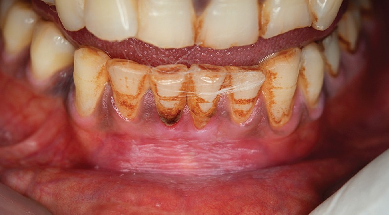
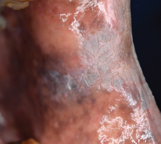
Tooth staining. These extrinsic stains are due to long term betel quid with tobacco use.
References
-
- National Cancer Institute and Centers for Disease Control and Prevention. Smokeless Tobacco and Public Health: A Global Perspective. NIH Publication No. 14-7983. 2014
-
- Richter P, Hodge K, Stanfill S, Zhang L, Watson C. Surveillance of moist snuff: Total nicotine, moisture, pH, un-ionized nicotine, and tobacco-specific nitrosamines. Nicotine Tob Res. 2008;10:1645–52. - PubMed
-
- Stepanov I, Hecht SS, Ramakrishnan S, Gupta PC. Tobacco-specific nitrosamines in smokeless tobacco products marketed in India. Int J Cancer. 2005;116:16–9. - PubMed
-
- Arain SS, Kazi TG, Afridi HI, Talpur FN, Kazi AG, Brahman KD, et al. Correlation of arsenic levels in smokeless tobacco products and biological samples of oral cancer patients and control consumers. Biol Trace Elem Res. 2015;168:287–95. - PubMed
-
- Chaturvedi P. Resource Centre for Tobacco Free India. Available from: www.rctfi.org/, accessed on May 2, 2013 .
Publication types
MeSH terms
LinkOut - more resources
Full Text Sources
Other Literature Sources
Medical