. 2019 Jan;111(1):96-117.
doi: 10.1111/mmi.14141. Epub 2018 Oct 21.

Two pKM101-encoded proteins, the pilus-tip protein TraC and Pep, assemble on the Escherichia coli cell surface as adhesins required for efficient conjugative DNA transfer

Affiliations

Two pKM101-encoded proteins, the pilus-tip protein TraC and Pep, assemble on the Escherichia coli cell surface as adhesins required for efficient conjugative DNA transfer

Christian González-Rivera et al. Mol Microbiol. 2019 Jan.

Abstract

Mobile genetic elements (MGEs) encode type IV secretion systems (T4SSs) known as conjugation machines for their transmission between bacterial cells. Conjugation machines are composed of an envelope-spanning translocation channel, and those functioning in Gram-negative species additionally elaborate an extracellular pilus to initiate donor-recipient cell contacts. We report that pKM101, a self-transmissible MGE functioning in the Enterobacteriaceae, has evolved a second target cell attachment mechanism. Two pKM101-encoded proteins, the pilus-tip adhesin TraC and a protein termed Pep, are exported to the cell surface where they interact and also form higher order complexes appearing as distinct foci or patches around the cell envelope. Surface-displayed TraC and Pep are required for an efficient conjugative transfer, 'extracellular complementation' potentially involving intercellular protein transfer, and activation of a Pseudomonas aeruginosa type VI secretion system. Both proteins are also required for bacteriophage PRD1 infection. TraC and Pep are exported across the outer membrane by a mechanism potentially involving the β-barrel assembly machinery. The pKM101 T4SS, thus, deploys alternative routing pathways for the delivery of TraC to the pilus tip or both TraC and Pep to the cell surface. We propose that T4SS-encoded, pilus-independent attachment mechanisms maximize the probability of MGE propagation and might be widespread among this translocation superfamily.


Figures

Fig. 1.
Fig. 1.
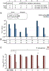
TraC and Pep stimulate intercellular contacts and mediate phage infection. Top: Strains used for the assays presented in panels A, B, and C. E. coli DH5α harbored the nontransmissible plasmids denoted in columns 1 – 8 that encode the TrapKM101 T4SS operon with (+) or deleted (Δ) of traC or pep;: 1. pKM101, 2. pCGR125, 3. pCGR135, 4. pCGR107, 5. pJG127, 6. pJG127 + pDA105; 7. pJG127 + pMB006; 8. pJJG127 + pMB007. Δ(c-traC, c-pep, or c-orf70): the complemented strain expresses the gene listed from the PnahG promoter. A) Conjugation assays. Donor strains additionally harbored the transmissible plasmid poriT (pJG142), which carries pKM101’s oriT sequence and traI (relaxase), traK (accessory factor), and traJ (T4CP) mobilization genes. Donors were mated with the plasmid-free recipient (MC4100-Rif) for 2-h in liquid or on a nitrocellulose disc on LB plates (Solid-surface). Transfer frequencies depicted in the bar graph are presented as transconjugants (Tc’s)/donor. B) PRD1 and IKe phage sensitivity of E. coli strains carrying the above-listed plasmids; (S) sensitive or (R) to infection, as monitored with a plaque assay. C) Survival of E. coli strains with the above-listed plasmids when cultivated in the absence or presence of P. aeruginosa PAO1. Statistical significance is shown based on a Student’s t test corresponding to the values of plasmid-free DH5α or growth in the absence of P. aeruginosa (*P<0.05). Data presented are mean ±SE, n=3 independent replicates.
Fig. 2.
Fig. 2.
TraC and Pep localize on the E. coli cell surface. A) Upper: TraCST is surface-displayed in the absence of the TrapKM101 T4SS as determined with a dot blot assay. Strains: MC4100 producing the indicated proteins from plasmids in parantheses: TraCST (pCGR83), STTraC (pCGR66), MBPST (pCGR84), TraFST (pCGR48), untagged (WT) TraC (pCGR35). Whole cells were spotted onto nitrocellulose and probed for detection of Strep-tagged proteins (with α-Strep), surface-exposed RcsF lipoprotein (α-RcsF), or the periplasmic POTRA domains of BamA (α-BamA POTRA). Lower: Lysates from cells deposited onto the nitrocellulose were analyzed for steady-state accumulation of Strep-tagged TraC, MBP, and TraF by SDS-PAGE and immunostaining with α-Strep antibodies. Left: Molecular size markers (kDa). Blots were also developed with antibodies to the RNAP β subunit as a loading control. B) TraC.V144CST reacts with BSA-Mal in the absence of the Tra pKM101 T4SS. Strains: MC4100 producing TraCST (pDA107), TraC.Q46CST (pDA110), or TraC.V144CST (pDA112). Intact cells were treated (+) or not treated (−) with BSA-Mal, the reactions were quenched, and cell lysates were analyzed for formation of TraCST/BSA-Mal complexes by SDS-PAGE and immunostaining with α-FLAG antibodies. Numbers at left: molecular size markers (kDa). Positions of higher-order TraCST/BSA-Mal complexes and monomeric TraCST are indicated. C) Identification of PepFL in the extracellular fraction obtained by shearing of cells. Strains: MC4100 cells producing PepFL (from pDA103), native Pep (pDA108), or MBPFL (pDA106). Whole cell extracts (left) or extracellular material recovered by shearing of cells (right) was analyzed for the presence of PepFL, and periplasmic MBPFL as a control, by immunostaining with α-FLAG antibodies. A nonspecific crossreactive species detected in extracellular fractions from all strains served as a loading control. Immunoblots were also stained with antibodies reactive to the periplasmic POTRA domains of BamA and the cytosolic β subunit of RNA polymerase (RNAP). D) Pep is displayed on the cell surface in the dot blot assay. Strains: MC4100 producing PepFL, Pep, or MBPFL from the above-listed plasmids, or Pep bearing FLAG tags immediately following the residue indicated from plasmids pDA113, 117, 115, 119, 116, 120, or 121. Whole cells were spotted onto nitrocellulose and probed with α-FLAG for detection of FLAG-tagged proteins, α-RcsF for surface-exposed RcsF lipoprotein, or α-BamA POTRA for periplasmic POTRA domains of BamA. Lysates from cells deposited onto the nitrocellulose were analyzed for PepFL or MBPFL protein abundance by SDS-PAGE and immunostaining. Left: Molecular size markers (kDa). Blots were probed with α-RNAP for detection of cytosolic β-subunit of RNA polymerase as a loading control. E) PepC105FL crosslinks with membrane-impermeable BSA-Mal. Strains: MC4100 producing PepFL (pDA103) or Pep.C105FL (pMB005). Intact cells were treated (+) or not treated (−) with BSA-Mal, the reactions were quenched, and cell lysates were analyzed for formation of PepFL/BSA-Mal complexes by SDS-PAGE and immunostaining with α-FLAG antibodies. Right: Positions of higher-order PepFL/BSA-Mal complexes and monomeric PepFL are indicated. Left: Molecular size markers (kDa).
Fig. 3.
Fig. 3.
TraC and Pep form punctate foci or patches on the cell surface. A) Left: Strain MC4100(pCGR110) producing TraCST in the presence of the TrapKM101 T4SS. TraCST puncta were detected by reactivity with α-Strep rabbit primary and AlexaFluor 594-conjugated goat α-rabbit secondary antibody. Cells were visualized by staining with Hoechst 3322 (blue, right panel), or the Hoechst emission channel was omitted (unstained cells, right panel). Right: Strain MC4100(pCGR127) producing PepFL from the kikA locus in the presence of the TrapKM101 T4SS. PepFL puncta and patches were visualized by reactivity with α-FLAG primary and AlexaFluor 488-conjugated secondary antibody, and cells by Hoechst staining as described above. B) Left and Right: MC4100 strains producing TraCST (from pCGR83) or PepFL (pDA103) in the absence of the TrapKM101 T4SS. TraCST, PepFL and cells were stained as described above. Scale bar = 5 μm. 3D-SIM localization patterns of TraCST and PepFL were obtained using a Nikon n-SIM Structured Illumination Super Resolution Microscope System housed at the Nikon Center of Excellence at UTHealth in the McGovern Medical School.
Fig. 4.
Fig. 4.
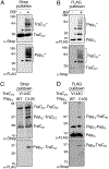
Extracellular TraC and Pep interact and form higher-order complexes. A and B) Whole cells of strain MC4100(pMB006), which coproduces TraCST and PepFL, were untreated (−) or treated (+) with BS3 crosslinker, and extracellular material was subjected to affinity pulldowns. Immunoblots were analyzed as indicated with α-Strep for detection of TraCST complexes or α-FLAG for detection of PepFL complexes. Monomeric forms of proteins are identified as TraCST or PepFL, crosslinked higher-order species are identified as TraCST* or PepFL*. Putative TraCST-PepFL heterodimers are denoted with black arrow heads, putative PepFL homodimers are denoted with a white arrow head. C and D) Cys-substituted variants of TraC and Pep form disulfide crosslinked homodimers. Strains: MC4100(pDA112) coproduces TraC.V144CST (V144C) and PepFL (WT); MC4100(pDA111) coproduces TraC.V144CST and Pep.C105FL. Material from the pull-downs was analyzed by nonreducing SDS-PAGE and immunoblot development with α-Strep or α-FLAG antibodies for detection of PepFL or TraCST complexes, respectively. Monomeric TraCST and PepFL and homo- or heterodimeric complexes are indicated. Left: Molecular size markers (kDa).
Fig. 5.
Fig. 5.
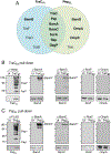
TraCST and PepFL interact with components of the BAM pathway. Strain MC4100(pCGR129) produces the TrapKM101 T4SS plus TraCST and PepFL from the tra locus. A) TraCST- and PepFL-interacting proteins were identified by affinity pull-downs and high-resolution nano-flow LC/MS. Venn Diagram shows proteins recovered in TraCST or PepFL pulldowns, or with both sets of pulldowns. Quantitative data for this strain and the control strain MC4100(pCGR125) producing untagged forms of TraC and Pep are presented in Table S1. B and C) Detection of BS3-crosslinked proteins in material recovered from TraCST (B) or PepFL (C) pull-downs. Strains: MC4100(pCGR129) coproduces TraCST and PepFL; MC4100(pCGR125) (denoted Ut) produces untagged forms of TraC and Pep. Material recovered from both sets of pull-downs was analyzed by immunoblotting for TraCST with α-Strep, PepFL with α-FLAG, or BamA, BamC, RcsF, or OmpA with respective antibodies. Molecular size markers (in kDa) at left. Lower panels: Total cell extracts from MC4100 carrying pCGR125 or pCGR129 were analyzed for the presence of each protein indicated. Higher-order crosslinked species of TraCST, PepFL BamA, and BamC are identified with an asterisk.
Fig. 6.
Fig. 6.
Evidence for involvement of the BAM pathway for export of TraC and Pep. A) Photocrosslinking of pBPA-substituted TraCST variants with BamA. MC4100 strains produced TraCST (from pCGR83), or TraCST variants with pBPA incorporated at positions Q46 (pCGR86), D73 (pCGR87), L86 (pCGR88), Q191 (pCGR93), or A218 (pCGR94). Cells were treated with UV and isolated membrane fractions were analyzed for the presence of TraCST-BamA complexes by immunostaining with α-BamA antibodies (top panel) or total pBPA-crosslinked TraCST species (lower panel) by immunostaining with α-Strep antibodies. B) pBPA substitutions at the positions indicated in a space-filled model of the TraC X-ray structure (Yeo et al., 2003) formed detectable photocrosslinks with BamA. C and D) Effects of BAM pathway mutations on accumulation of extracellular complexes of TraCST and PepFL. Strains: MC4100 or indicated mutant strains producing TraCST from pDA107 (panel C) or PepFL from pDA103 (panel D). Cells were treated with BS3 (+BS3) and extracellular fractions were subjected to α-Strep or pull-downs. Material recovered from the pull-downs was analyzed for the presence of higher-order TraCST or PepFL by immunostaining. Monomeric and higher-order complexes (denoted with *) of TraCST and PepFL are indicated. Monomeric PepFL was detected upon overexposure (o.e.) of immunoblot (D, lower panel). Numbers below top panels correspond to relative abundance of crosslinked species compared with levels detected in MC4100(pDA107 or pDA103) cells (lane 1, set to 1), as determined by densitometry tracing of the regions of each lane demarked with] at the right of the panel using Image J software. Bottom panel: Total cellular levels (-BS3) of TraCST and PepFL in whole cell extracts of the strains listed at the top. A, C, & D) Molecular size markers (in kDa) at left.

Similar articles

Cited by

References

    1. Achtman M, (1975) Mating aggregates in Escherichia coli conjugation. J Bacteriol 123: 505–515. - PMC - PubMed
    1. Achtman M, Morelli G, and Schwuchow S (1978) Cell-cell interactions in conjugating Escherichia coli: role of F pili and fate of mating aggregates. J Bacteriol 135: 1053–1061. - PMC - PubMed
    1. Alvarez-Martinez CE, and Christie PJ (2009) Biological diversity of prokaryotic type IV secretion systems. Microbiol Mol Biol Rev 73: 775–808. - PMC - PubMed
    1. Aly KA, and Baron C (2007) The VirB5 protein localizes to the T-pilus tips in Agrobacterium tumefaciens. Microbiology 153: 3766–3775. - PubMed
    1. Anderson LB, Hertzel AV, and Das A (1996) Agrobacterium tumefaciens VirB7 and VirB9 form a disulfide-linked protein complex. Proc Natl Acad Sci USA 93: 8889–8894. - PMC - PubMed

Publication types

MeSH terms