. 2018 Nov 23;293(47):18230-18241.
doi: 10.1074/jbc.RA118.004187. Epub 2018 Sep 28.

Angiomotins stimulate LATS kinase autophosphorylation and act as scaffolds that promote Hippo signaling

Affiliations

Angiomotins stimulate LATS kinase autophosphorylation and act as scaffolds that promote Hippo signaling

Sebastian Mana-Capelli et al. J Biol Chem. .

Abstract

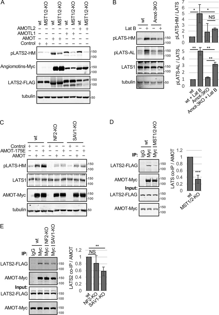
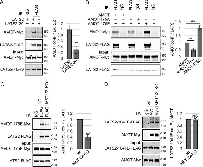
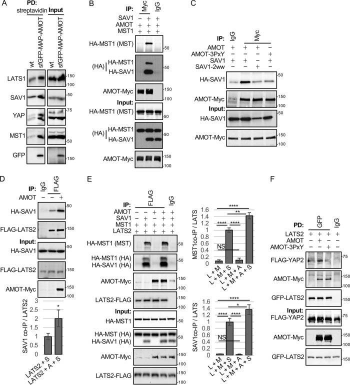
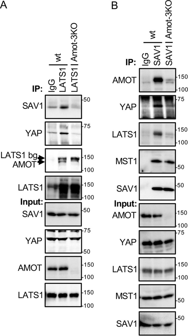
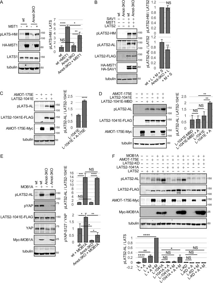
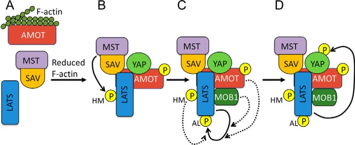
The Hippo pathway controls cell proliferation, differentiation, and survival by regulating the Yes-associated protein (YAP) transcriptional coactivator in response to various stimuli, including the mechanical environment. The major YAP regulators are the LATS1/2 kinases, which phosphorylate and inhibit YAP. LATS1/2 are activated by phosphorylation on a hydrophobic motif (HM) outside of the kinase domain by MST1/2 and other kinases. Phosphorylation of the HM motif then triggers autophosphorylation of the kinase in the activation loop to fully activate the kinase, a process facilitated by MOB1. The angiomotin family of proteins (AMOT, AMOTL1, and AMOTL2) bind LATS1/2 and promote its kinase activity and YAP phosphorylation through an unknown mechanism. Here we show that angiomotins increase Hippo signaling through multiple mechanisms. We found that, by binding LATS1/2, SAV1, and YAP, angiomotins function as a scaffold that connects LATS1/2 to both its activator SAV1-MST1 and its target YAP. Deletion of all three angiomotins reduced the association of LATS1 with SAV1-MST1 and decreased MST1/2-mediated LATS1/2-HM phosphorylation. Angiomotin deletion also reduced LATS1/2's ability to associate with and phosphorylate YAP. In addition, we found that angiomotins have an unexpected function along with MOB1 to promote autophosphorylation of LATS1/2 on the activation loop motif independent of HM phosphorylation. These results indicate that angiomotins enhance Hippo signaling by stimulating LATS1/2 autophosphorylation and by connecting LATS1/2 with both its activator SAV1-MST1/2 and its substrate YAP.

Keywords: AMOT; Hippo pathway; LATS (Warts, Wts); MOB1; SAV1; Salvador (Sav); Yes-associated protein (YAP); angiomotin; cell signaling; mammalian sterile 20-like kinase 1 (MST1); mammalian sterile 20-like kinase 2 (MST2); mechanotransduction; protein kinase; scaffold protein.


Conflict of interest statement

The authors declare that they have no conflicts of interest with the contents of this article. The content is solely the responsibility of the authors and does not necessarily represent the official views of the National Institutes of Health

Figures

Figure 1.
Figure 1.
Angiomotins require MST1/2, SAV1, and NF2 to activate LATS1/2. A, HEK293 (WT) or HEK293 cells with MST1 and MST2 inactivated using CRISPR (MST1/2-KO) were transfected with a LATS2-FLAG–expressing plasmid and either a control plasmid or a plasmid for expressing Myc-tagged versions of each of the three angiomotin genes (AMOT, AMOTL1, and AMOTL2). Cell lysates were analyzed by Western blotting using antibodies against the LATS1/2-HM phosphorylation site (pLATS2-HM), Myc (Angiomotins-Myc), and FLAG (LATS2-FLAG). Tubulin levels in cell lysates are also shown. B, HEK293 (WT) or HEK293 cells with AMOT, AMOTL1, and AMOTL2 inactivated using CRISPR (Amot-3KO) were treated with or without Lat B, and cell lysates were analyzed by Western blotting using antibodies against LATS1/2-HM phosphorylation, LATS1/2-AL phosphorylation, and LATS1. Tubulin levels in cell lysates are also shown. Quantification of LATS1/2-HM and LATS1/2-AL phosphorylation relative to untreated WT HEK293 cells is shown. Mean ± S.D.; n = 3; NS, p ≥ 0.05; *, p ≤ 0.05; **, p ≤ 0.01; t test. C, HEK293 (WT) or HEK293 cells with NF2 (NF2-KO) or SAV1 (SAV1-KO) inactivated using CRISPR were transfected with a control plasmid or a plasmid for expressing Myc-tagged versions of AMOT or the AMOT-S175E mutant. Cell lysates were analyzed by Western blotting using antibodies against the LATS1/2-HM phosphorylation site (pLATS1/2-HM) and Myc (AMOT-Myc). Tubulin levels in cell lysates are also shown. D, HEK293 (WT) or MST1/2-KO HEK293 cells were transfected with LATS2-FLAG– and AMOT-Myc–expressing plasmids. Myc or control (IgG) antibodies were used for immunoprecipitation (IP) from cell lysates, and immune complexes and cell lysates were analyzed by Western blotting for LATS2-FLAG and AMOT-Myc levels. Quantification of LATS2 levels in AMOT immune complexes is shown. Mean ± S.D.; n = 3; ***, p ≤ 0.001; t test. E, HEK293 (WT), NF2-KO, or SAV1-KO HEK293 cells were transfected with LATS2-FLAG and AMOT-Myc expressing plasmids. Myc or control (IgG) antibodies were used for immunoprecipitations from cell lysates, and immune complexes and cell lysates were analyzed by Western blotting for LATS2-FLAG and AMOT-Myc levels. Quantification of LATS2 levels in AMOT immune complexes is shown. Mean ± S.D.; n = 3; **, p ≤ 0.01; NS, p ≥ 0.05; t test.
Figure 2.
Figure 2.
AMOT binds to active forms of LATS2. A, HEK293 cells were transfected with plasmids for expressing AMOT-Myc and either LATS2 or LATS2 with the AL and HM phosphorylation sites (Ser872 and Thr1041) mutated to alanine (LATS2–2A). Myc or control (IgG) antibodies were used for immunoprecipitation (IP) from cell lysates, and immune complexes and cell lysates were analyzed by Western blotting for LATS2-FLAG and AMOT-Myc levels. Quantification of AMOT levels in LATS2 immune complexes is shown. Mean ± S.D.; n = 3; **, p ≤ 0.01; t test). B, HEK293 cells were transfected with plasmids for expressing LATS2-FLAG and either AMOT-Myc, AMOT-175A-Myc, or AMOT-175E-Myc. Immunoprecipitations were performed on each cell lysate with either FLAG (LATS2-FLAG) or control (IgG) antibodies, and immune complexes and cell lysates were analyzed by Western blotting for LATS2-FLAG and AMOT-Myc levels. Quantification of AMOT levels in LATS2 immune complexes is shown. Mean ± S.D.; n = 3; **, p ≤ 0.01; ***, p ≤ 0.001; t test. C, HEK293 cells (WT) or MST1/2-KO cells were transfected with plasmids for expressing LATS2-FLAG and AMOT-175E-Myc. Immunoprecipitations were performed on each cell lysate with either FLAG (LATS2-FLAG) or control (IgG) antibodies, and immune complexes and cell lysates were analyzed by Western blotting for LATS2-FLAG and AMOT-Myc levels. Quantification of AMOT levels in LATS2 immune complexes is shown. Mean ± S.D.; n = 3; ***, p ≤ 0.001; t test. D, HEK293 cells (WT) or MST1/2-KO cells were transfected with plasmids for expressing LATS2-T1041E-FLAG and AMOT-Myc. Immunoprecipitations were performed on cell lysates with either Myc (AMOT-Myc) or control (IgG) antibodies, and immune complexes and cell lysates were analyzed by Western blotting for LATS2-FLAG and AMOT-Myc levels. Quantification of LATS2 levels in AMOT immune complexes is shown. Mean ± S.D.; n = 3; NS, p ≥ 0.05; t test.
Figure 3.
Figure 3.
AMOT promotes assembly of LATS2-SAV1-MST1 complexes. A, HEK293 cells expressing sfGFP-AMOT from the chromosomal locus were created using CRISPR-mediated genome modification (see “Experimental procedures”). Streptavidin beads were used to pull down (PD) sfGFP-MAP-AMOT protein (GFP-AMOT) (WT HEK293 cells where the endogenous AMOT locus has not been tagged with the sfGFP-MAP tag were used as a control). Protein complexes on beads were analyzed by Western blotting using antibodies against the indicated proteins. The levels of these proteins in cell lysates are shown. B, HEK293 cells were transfected with plasmids for expressing HA-MST1, HA-SAV1, and AMOT-Myc as indicated. Immunoprecipitation (IP) was performed cell lysates with either Myc (AMOT-Myc) or control (IgG) antibodies, and immune complexes and cell lysates were analyzed by Western blotting for HA-MST1, HA-SAV1, and AMOT-Myc levels. C, HEK293 cells were transfected with plasmids for expressing HA-SAV1 or HA-SAV1 with both WW domains mutated (HA-SAV1–2ww) and either AMOT-Myc or AMOT-3PxY-Myc (eliminates all three L/PPXY motifs in AMOT). Immunoprecipitations were performed on each cell lysate with either Myc (AMOT-Myc) or control (IgG) antibodies, and immune complexes and cell lysates were analyzed by Western blotting for HA-SAV1 and AMOT-Myc levels. D, HEK293 cells were transfected with plasmids for expressing HA-SAV1, LATS2-FLAG, and AMOT-Myc as indicated. Immunoprecipitations were performed on each cell lysate with either FLAG (LATS2-FLAG) or control (IgG) antibodies, and immune complexes and cell lysates were analyzed by Western blotting for LATS2-FLAG, HA-SAV1, and AMOT-Myc levels. Quantification of SAV/LATS2 ratios in LATS2 immune complexes for cells expressing LATS2 and SAV1 (LATS2+S) or LATS2, SAV1, and AMOT (LATS2+A+S) is shown. Mean ± S.D.; n = 3; *, p ≤ 0.05; t test). E, HEK293 cells were transfected with combinations of plasmids for expressing LATS2-FLAG, HA-MST1, HA-SAV1, and AMOT-Myc as indicated. Immunoprecipitations were performed on each cell lysate with either FLAG (LATS2-FLAG) or control (IgG) antibodies, and immune complexes and cell lysates were analyzed by Western blotting for LATS2-FLAG, HA-MST1, HA-SAV1, and AMOT-Myc levels. Quantification of MST1/LATS2 and SAV1/LATS2 ratios in LATS2 immune complexes for cells co-expressing LATS2 and MST1 (L + M); LATS2, MST1, and SAV (L + M + S); LATS2, MST1, and AMOT (L + M + A); and LATS2, MST1, AMOT, and SAV1 (L + M + A + S) is shown. Mean ± S.D.; n = 3; *, p ≤ 0.05; **, p ≤ 0.01; ****, p ≤ 0.0001; t test. NS, not significant. F, HEK293 cells were transfected with combinations of plasmids for expressing GFP-LATS2, FLAG-YAP2, and AMOT-Myc as indicated. Immunoprecipitations were performed on each cell lysate with either GFP (LATS2-GFP) or control (IgG) antibodies, and immune complexes and cell lysates were analyzed by Western blotting for LATS2-GFP, FLAG-YAP2, and AMOT-Myc levels.
Figure 4.
Figure 4.
Angiomotins function as scaffolds for the Hippo pathway in vivo. A, HEK293 (WT) and Amot-3KO HEK293 cells were grown to high density and serum-starved for 4 h. Immunoprecipitation (IP) was performed on cell lysates with either anti-LATS1 or control (IgG) antibodies, and immune complexes and cell lysates were analyzed by Western blotting for AMOT, SAV1, LATS1, and YAP levels. LATS1 bg, residual LATS1 signal after stripping. B, HEK293 (WT) and Amot-3KO HEK293 cells were grown as in A. Immunoprecipitations were performed on cell lysates with either anti-SAV1 or control (IgG) antibodies, and immune complexes and cell lysates were analyzed by Western blotting for AMOT, MST1, SAV1, LATS1, and YAP levels.
Figure 5.
Figure 5.
AMOT promotes LATS2-AL activation loop phosphorylation independent of LATS2-HM phosphorylation. A, HEK293 (WT) or Amot-3KO HEK293 cells were transfected with a control plasmid or a plasmid for expressing HA-MST1. Cell lysates were analyzed by Western blotting using antibodies against the LATS1/2-HM phosphorylation site (pLATS1/2-HM), HA (HA-MST1), and LATS1. Tubulin levels in cell lysates are also shown. Quantification of LATS1/2-HM phosphorylation is shown. Mean ± S.D.; n = 3; NS, p ≥ 0.05; *, p ≤ 0.05; **, p ≤ 0.01; ****, p ≤ 0.0001; t test. B, HEK293 (WT) or Amot-3KO KO HEK293 cells were transfected with a control plasmid or a combination of plasmids (L + M + S) for expressing LATS2-FLAG, HA-MST1, and HA-SAV1. Cell lysates were analyzed by Western blotting using antibodies for LATS2-HM and LATS2-AL phosphorylation, FLAG (LATS2-FLAG), and HA (HA-MST1 and HA-SAV1). Tubulin levels in cell lysates are also shown. Quantification of LATS1/2-HM phosphorylation is shown. Mean ± S.D.; n = 3; NS, p ≥ 0.05; **, p ≤ 0.001; t test. C, HEK293 cells were transfected with a control plasmid, LATS2–1041E-FLAG, or AMOT-175E-Myc as indicated. Cell lysates were analyzed by Western blotting using antibodies for LATS2-AL phosphorylation (pLATS2-AL), FLAG (LATS2–1041E-FLAG), and Myc (AMOT-175E-Myc). Tubulin levels in cell lysates are also shown. Quantification of LATS1/2-AL phosphorylation relative to LATS2–1041E is shown. L-1041E, LATS2–1041E; A-175E, AMOT-175E. Mean ± S.D.; n = 3; ****, p ≤ 0.0001; t test. D, HEK293 cells were transfected with a control plasmid or LATS2–1041E-FLAG, LATS2–1041E-MBD-FLAG (MBD is a LATS2 mutant defective for binding MOB1), or AMOT-175E-Myc as indicated. Cell lysates were analyzed by Western blotting using antibodies for LATS2-AL phosphorylation (pLATS2-AL), FLAG (LATS2–1041E-FLAG or LATS2–1041E-MBD-FLAG), and Myc (AMOT-Myc). Tubulin levels in cell lysates are also shown. Quantification of LATS1/2-AL phosphorylation relative to LATS2–1041E-FLAG levels is shown. L-1041E, LATS2–1041E; A, AMOT-175E. Mean ± S.D.; n = 3; NS, p ≥ 0.05; **, p ≤ 0.01; t test). E, HEK293 (WT) or Amot-3KO HEK293 cells were transfected with a plasmid for expressing LATS2–1041E-FLAG and either a control plasmid or a plasmid for expressing Myc-MOB1A. Cell lysates were analyzed by Western blotting using antibodies for LATS2-AL phosphorylation (pLATS2-AL), YAP phosphorylation on Ser127 (pYAP-S127), FLAG (LATS2–1041E-FLAG), and Myc (Myc-MOB1A). Tubulin levels in cell lysates are also shown. Quantification of LATS1/2-AL and pYAP-Ser127 phosphorylation is shown. Mean ± S.D.; n = 3; NS, p ≥ 0.05; *, p ≤ 0.05; **, p ≤ 0.01; ***, p ≤ 0.001; ****, p ≤ 0.0001; t test). F, HEK293 cells were transfected with a control plasmid, LATS2-FLAG, LATS2–1041E-FLAG, kinase-dead LATS2 (LATS2-KD-FLAG), AMOT-Myc, or Myc-MOB1A as indicated. Cell lysates were analyzed by Western blotting using antibodies for LATS2-AL phosphorylation (pLATS2-AL), FLAG (LATS2-FLAG), or Myc (AMOT-Myc and Myc-MOB1A). Tubulin levels in cell lysates are also shown. L, LATS2; L-1041A, LATS2–1041A; L-KD, LATS2-kinase dead; A, AMOT-175E; M, MOB1A. Quantification of LATS1/2-AL phosphorylation is shown. Mean ± S.D.; n = 3; NS, p ≥ 0.05; *, p ≤ 0.05; **, p ≤ 0.01; ****, p ≤ 0.0001; t test).
Figure 6.
Figure 6.
Model for AMOT function in Hippo signaling. A, when F-actin levels are high, AMOT is bound to F-actin and interacts poorly with its binding partners. B, when F-actin levels are reduced, AMOT becomes phosphorylated by LATS and is free to bind YAP, SAV-MST, and LATS. Assembly of this complex allows MST to phosphorylate LATS on the HM site. Both AMOT phosphorylation and LATS HM phosphorylation enhance AMOT-LATS binding. C, to become fully active, LATS must autophosphorylate on the AL site. HM phosphorylation enhances LATS autophosphorylation. In addition, we show that both MOB1 and AMOT enhance LATS-AL phosphorylation independent of any effects they have on HM phosphorylation. D, the ability of fully active LATS to phosphorylate YAP may be enhanced by formation of a LATS–AMOT–YAP complex. Note that the model is speculative and its purpose is to illustrate the primary steps involved in AMOT activation of Hippo signaling. The Hippo pathway is regulated at many other levels, which could be occurring at the same time but are not shown for simplicity. It is presently not clear whether complexes exist in cells as pictured, with all components simultaneously bound to each other.

Similar articles

Cited by

References

    1. Meng Z., Moroishi T., and Guan K. L. (2016) Mechanisms of Hippo pathway regulation. Genes Dev. 30, 1–17 10.1101/gad.274027.115 - DOI - PMC - PubMed
    1. Zanconato F., Cordenonsi M., and Piccolo S. (2016) YAP/TAZ at the roots of cancer. Cancer Cell 29, 783–803 10.1016/j.ccell.2016.05.005 - DOI - PMC - PubMed
    1. Dong J., Feldmann G., Huang J., Wu S., Zhang N., Comerford S. A., Gayyed M. F., Anders R. A., Maitra A., and Pan D. (2007) Elucidation of a universal size-control mechanism in Drosophila and mammals. Cell 130, 1120–1133 10.1016/j.cell.2007.07.019 - DOI - PMC - PubMed
    1. Zhao B., Li L., Tumaneng K., Wang C. Y., and Guan K. L. (2010) A coordinated phosphorylation by Lats and CK1 regulates YAP stability through SCF(β-TRCP). Genes Dev. 24, 72–85 10.1101/gad.1843810 - DOI - PMC - PubMed
    1. Liu C. Y., Zha Z. Y., Zhou X., Zhang H., Huang W., Zhao D., Li T., Chan S. W., Lim C. J., Hong W., Zhao S., Xiong Y., Lei Q. Y., and Guan K. L. (2010) The hippo tumor pathway promotes TAZ degradation by phosphorylating a phosphodegron and recruiting the SCFβ-TrCP E3 ligase. J. Biol. Chem. 285, 37159–37169 10.1074/jbc.M110.152942 - DOI - PMC - PubMed

Publication types

MeSH terms