. 2018 Nov 16;293(46):17863-17874.
doi: 10.1074/jbc.RA118.004548. Epub 2018 Sep 28.

Poly(ADP-ribose) polymerase 1 (PARP1) promotes oxidative stress-induced association of Cockayne syndrome group B protein with chromatin

Affiliations

Poly(ADP-ribose) polymerase 1 (PARP1) promotes oxidative stress-induced association of Cockayne syndrome group B protein with chromatin

Erica L Boetefuer et al. J Biol Chem. .

Abstract

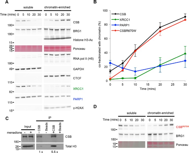
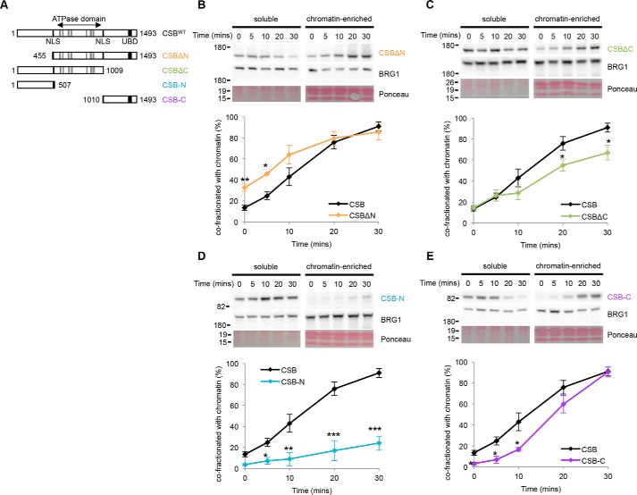
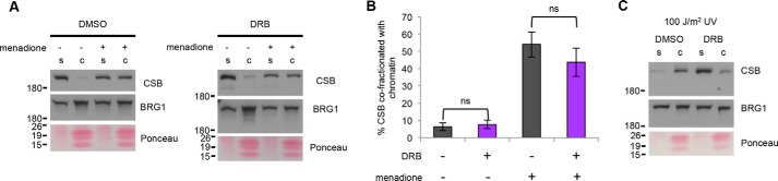
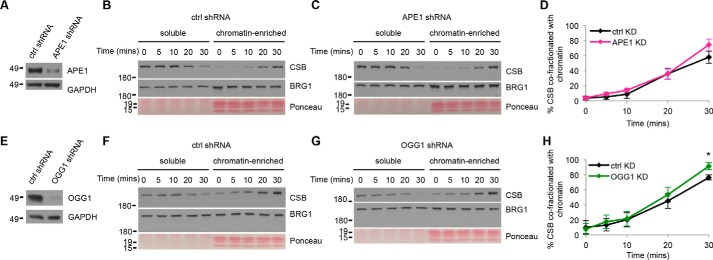
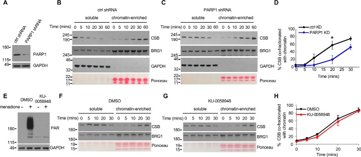
Cockayne syndrome protein B (CSB) is an ATP-dependent chromatin remodeler that relieves oxidative stress by regulating DNA repair and transcription. CSB is proposed to participate in base-excision repair (BER), the primary pathway for repairing oxidative DNA damage, but exactly how CSB participates in this process is unknown. It is also unclear whether CSB contributes to other repair pathways during oxidative stress. Here, using a patient-derived CS1AN-sv cell line, we examined how CSB is targeted to chromatin in response to menadione-induced oxidative stress, both globally and locus-specifically. We found that menadione-induced, global CSB-chromatin association does not require CSB's ATPase activity and is, therefore, mechanistically distinct from UV-induced CSB-chromatin association. Importantly, poly(ADP-ribose) polymerase 1 (PARP1) enhanced the kinetics of global menadione-induced CSB-chromatin association. We found that the major BER enzymes, 8-oxoguanine DNA glycosylase (OGG1) and apurinic/apyrimidinic endodeoxyribonuclease 1 (APE1), do not influence this association. Additionally, the level of γ-H2A histone family member X (γ-H2AX), a marker for dsDNA breaks, was not increased in menadione-treated cells. Therefore, our results support a model whereby PARP1 localizes to ssDNA breaks and recruits CSB to participate in DNA repair. Furthermore, this global CSB-chromatin association occurred independently of RNA polymerase II-mediated transcription elongation. However, unlike global CSB-chromatin association, both PARP1 knockdown and inhibition of transcription elongation interfered with menadione-induced CSB recruitment to specific genomic regions. This observation supports the hypothesis that CSB is also targeted to specific genomic loci to participate in transcriptional regulation in response to oxidative stress.

Keywords: ATP-dependent chromatin remodeling; CSB; Cockayne syndrome protein; DNA repair; PARP1; base-excision repair (BER); chromatin; oxidative stress; reactive oxygen species (ROS); single-strand DNA repair; transcription regulation.


Conflict of interest statement

The authors declare that they have no conflicts of interest with the contents of this article

Figures

Figure 1.
Figure 1.
The association of CSB with chromatin in response to menadione treatment occurs independently of ATP hydrolysis. A, protein fractionation assay in CS1AN-CSBWT cells following treatment with 100 μm menadione for times indicated. Western blots were probed with antibodies listed. BRG1 was used as a loading control. Acetylated histone H3 and total core histones (visualized by Ponceau S staining) were used as markers for the chromatin-enriched fraction. GAPDH was used as a marker for the soluble fraction. B, quantification of percent CSBWT (n = 5), PARP1 (n = 4), XRCC1 (n = 4), and CSBR670W (n = 2) in the chromatin-enriched fraction as a function of time, normalized to BRG1. Error bars represent S.E. C, CSB ChIP-Western blot analysis in CS1AN-CSBWT cells untreated (−) or treated with 100 μm menadione for 30 min (+). IP, immunoprecipitation. Numbers at the bottom show -fold change in histone H3 normalized to CSB (n = 2). D, protein fractionation assay in CS1AN-CSBR670W cells following treatment with 100 μm menadione for times indicated (n = 2). Shown is a representative Western blot probed with antibodies to CSB and BRG1 and stained with Ponceau S.
Figure 2.
Figure 2.
The association of CSB with chromatin in response to menadione treatment is largely mediated through its ATPase domain and C-terminal region. A, schematic representation of the CSB protein and CSB deletion constructs used in the protein fractionation assays. Gray boxes represent the seven conserved helicase motifs, thin black boxes represent the two putative nuclear localization signals (NLS), and the thick black box represents the ubiquitin-binding domain (UBD). B–E, protein fractionation assays demonstrating chromatin association as a function of time after 100 μm menadione treatment in CS1AN-sv cells reconstituted with the indicated CSB derivatives: CSBWT (n = 5) (from Fig. 1A) and CSBΔN (n = 2) (B), CSBΔC (n = 3) (C), CSB-N (n = 2) (D), and CSB-C (n = 4) (E). Shown are representative Western blots probed with the indicated antibodies and stained with Ponceau S for histones. Plots show quantification of the Western blot data with CSB signals normalized to BRG1 signals. Error bars represent S.E. Paired t tests compare CSB derivative enrichment to CSBWT (*, p ≤ 0.05; **, p ≤ 0.01; ***, p ≤ 0.001).
Figure 3.
Figure 3.
Inhibiting transcription elongation of RNA pol II by DRB does not alter menadione-induced CSB–chromatin association. A, protein fractionation assay in CS1AN-CSBWT cells. Cells were treated with 50 μm DRB or DMSO for 1 h followed by a 100 μm menadione treatment for 20 min. Shown are representative Western blot probed with antibodies listed. S, denotes soluble protein fraction; C, denotes chromatin-enriched protein fraction. B, quantification of CSB chromatin co-fractionation data in A normalized to BRG1. Shown are means ± S.E., and paired t test compares enrichment in cells with DMSO versus DRB treatment (n = 3, ns, not significant). C, protein fractionation assay in CS1AN-CSBWT cells treated with 50 μm DRB or DMSO for 1 h followed by 100 J/m2 UV irradiation. Cells were analyzed 1 h after UV treatment.
Figure 4.
Figure 4.
APE1 or OGG1 are dispensable for menadione-induced global CSB–chromatin association. A, representative Western blots revealing the extent of APE1 knockdown (average knockdown ∼72%, normalized to GAPDH). B and C, protein fractionation assays revealing CSB–chromatin association as a function of time after menadione treatment in CS1AN-CSBWT cells expressing a control (ctrl) or APE1 shRNA. Shown are representative Western blots probed with antibodies listed and stained with Ponceau S. D, quantification of data in B and C showing percent CSB co-fractionating with chromatin. Error bars represent S.E. Paired t test comparing CSB enrichment in control versus APE1 knockdown (n = 4) revealed no significant differences in association kinetics. E, representative Western blots revealing the extent of OGG1 knockdown (average knockdown ∼90%, normalized to GAPDH). F and G, protein fractionation assays revealing CSB–chromatin association as a function of time after menadione treatment in CS1AN-CSBWT cells expressing a control or OGG1 shRNA. Shown are representative Western blots probed with antibodies listed and stained with Ponceau S. H, quantification of data in F and G showing percent CSB co-fractionating with chromatin. Error bars represent S.E. Paired t test compares CSB enrichment in control to OGG1 knockdown (n = 4; *, p ≤ 0.05).
Figure 5.
Figure 5.
The PARP1 protein, but not its enzymatic activity, is required for efficient global CSB–chromatin association in response to menadione treatment. A, representative Western blots revealing the extent of PARP1 knockdown (average knockdown ∼89%, normalized to GAPDH). B and C, protein fractionation assays revealing CSB–chromatin association as a function of time after menadione treatment in CS1AN-CSBWT cells expressing a control (ctrl) or PARP1 shRNA. Shown are representative Western blots probed with antibodies listed and stained with Ponceau S (the loading ratio of soluble to chromatin is 1:2.2). D, quantification of data in B and C showing percent CSB co-fractionating with chromatin. Error bars represent S.E. Paired t test compares CSB enrichment in control versus PARP1 knockdown (n = 4; *, p ≤ 0.05). E, Western blots probed with an anti-PAR antibody demonstrating PARP1 inhibition by KU-0058948. F and G, protein fractionation assays of CS1AN-CSBWT cells treated with DMSO (vehicle control) or KU-0058948 followed by the addition 100 μm menadione for the indicated times. Shown are representative Western blots probed with antibodies listed and stained with Ponceau S (the loading ratio of soluble to chromatin is 1:1.25). H, quantification of data in F and G showing percent CSB co-fractionating with chromatin. Error bars represent S.E. Paired t test comparing CSB enrichment in DMSO- versus KU-0058948–treated cells (n = 5) revealed no significant difference.
Figure 6.
Figure 6.
PARP1 and active transcription contribute to menadione-induced CSB occupancy at specific genomic loci. Shown are four loci where CSB occupancy is significantly enhanced by oxidative stress (chrX-1, chrX-2, chr17-1, and chr19-2) and a control locus where CSB occupancy is not changed by oxidative stress (chr12-7). A, CSB ChIP-qPCR analyses of CS1AN-CSBWT cells expressing a control (ctrl) or PARP1 shRNA. Shown are means ± S.E. (n = 3). B, CSB ChIP-qPCR analyses as above except that cells were exposed to KU-0058948 (PARP1 i) or DMSO for 1 h prior to menadione treatment. Shown are means ± S.E. (n = 2). C, CSB ChIP-qPCR analyses of cells exposed to DRB or DMSO for 1 h prior to menadione treatment. Shown are means ± S.E. (n = 2). D, ChIP-qPCR analyses of CSB enrichment at specific genomic loci in cells without (mock) or with α-amanitin (aA) treatment prior to menadione treatment. Cells were treated with 1 mg/ml α-amanitin for 1 h prior to menadione treatment for 20 min. Shown are means ± S.E. (n = 2). Paired t tests compare CSB enrichment (*, p ≤ 0.05; **, p ≤ 0.01; ***, p ≤ 0.001).
Figure 7.
Figure 7.
Models for CSB functions during oxidative stress. A, ssDNA breaks generated by reactive oxygen species are recognized by PARP1. Localization of PARP1 to single-strand breaks facilitates the recruitment of CSB. CSB binds chromatin through its ATPase domain. Upon oxidative stress, PARP1 binds to the CSB N- and C-terminal regions; this interaction exposes a chromatin interaction surface in the C-terminal region of CSB that stabilizes CSB–chromatin association. CSB may function to make the chromatin landscape more permissible for DNA repair and/or to regulate repair-protein retention at sites of repair. B, menadione sensitivity assays. The chromatin remodeling–deficient CSBΔN1 derivative does not complement the menadione sensitivity of CS1AN-sv cells. Paired t tests compare CS1AN-CSBWT with CS1AN-CSBΔN1 (n = 5; *, p ≤ 0.05; ***, p ≤ 0.001). C, menadione-induced CSB occupancy at specific genomic loci depends on PARP1 (this study) and CTCF (20). These proteins may likely organize higher-order chromatin structure to mount a transcriptional response to oxidative stress.

Similar articles

Cited by

References

    1. Lehmann A. R. (1982) Three complementation groups in Cockayne syndrome. Mut. Res. 106, 347–356 10.1016/0027-5107(82)90115-4 - DOI - PubMed
    1. Nance M. A., and Berry S. A. (1992) Cockayne syndrome: Review of 140 cases. Am. J. Med. Genet. 42, 68–84 10.1002/ajmg.1320420115 - DOI - PubMed
    1. Troelstra C., van Gool A., de Wit J., Vermeulen W., Bootsma D., and Hoeijmakers J. H. (1992) ERCC6, a member of a subfamily of putative helicases, is involved in Cockayne's syndrome and preferential repair of active genes. Cell 71, 939–953 10.1016/0092-8674(92)90390-X - DOI - PubMed
    1. Eisen J. A., Sweder K. S., and Hanawalt P. C. (1995) Evolution of the SNF2 family of proteins: Subfamilies with distinct sequences and functions. Nucleic Acids Res. 23, 2715–2723 10.1093/nar/23.14.2715 - DOI - PMC - PubMed
    1. Citterio E., Van Den Boom V., Schnitzler G., Kanaar R., Bonte E., Kingston R. E., Hoeijmakers J. H., and Vermeulen W. (2000) ATP-dependent chromatin remodeling by the Cockayne syndrome B DNA repair-transcription-coupling factor. Mol. Cell. Biol. 20, 7643–7653 10.1128/MCB.20.20.7643-7653.2000 - DOI - PMC - PubMed

Publication types

MeSH terms