. 2018 Oct 9;2(19):2467-2477.
doi: 10.1182/bloodadvances.2018018309.

ASXL1 impairs osteoclast formation by epigenetic regulation of NFATc1

Affiliations

ASXL1 impairs osteoclast formation by epigenetic regulation of NFATc1

Nidhi Rohatgi et al. Blood Adv. .

Abstract

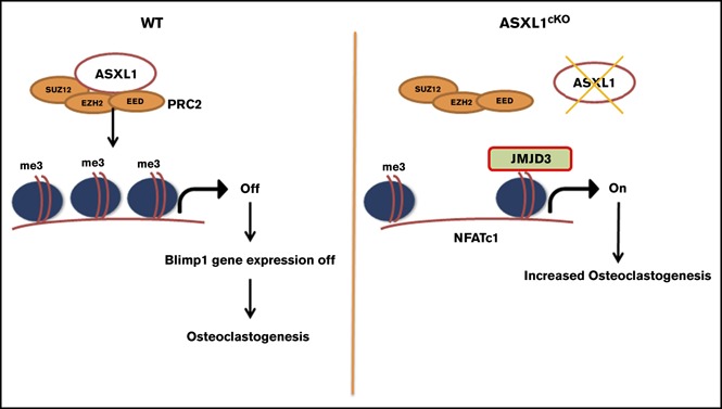
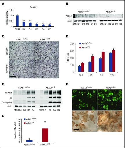
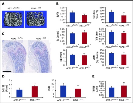
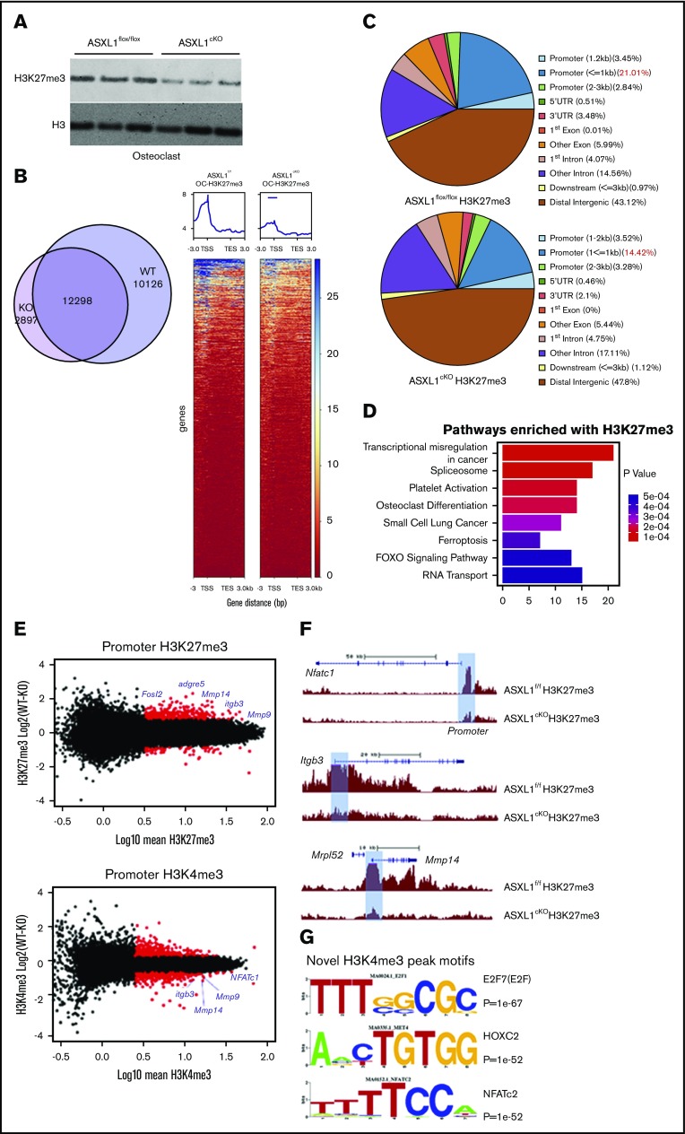
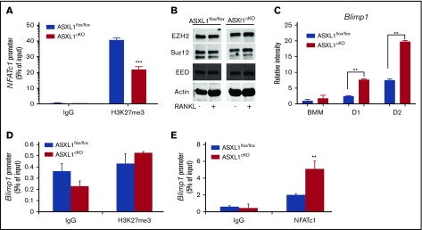
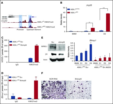
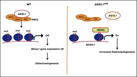
Additional sex comb-like 1 (ASXL1) mutations are commonly associated with myeloid malignancies and are markers of aggressive disease. The fact that ASXL1 is necessary for myeloid differentiation raises the possibility it also regulates osteoclasts. We find deletion of ASXL1 in myeloid cells results in bone loss with increased abundance of osteoclasts. Because ASXL1 is an enhancer of trithorax and polycomb (ETP) protein, we asked if it modulates osteoclast differentiation by maintaining balance between positive and negative epigenetic regulators. In fact, loss of ASXL1 induces concordant loss of inhibitory H3K27me3 with gain of H3K4me3 at key osteoclast differentiation genes, including nuclear factor for activated T cells 1 (NFATc1) and itgb3 In the setting of ASXL1 deficiency, increased NFATc1 binds to the Blimp1 (Prdm1) promoter thereby enhancing expression of this pro-osteoclastogenic gene. The global reduction of K27 trimethylation in ASXL1-deficient osteoclasts is also attended by a 40-fold increase in expression of the histone demethylase Jumonji domain-containing 3 (Jmjd3). Jmjd3 knockdown in ASXL1-deficient osteoclast precursors increases H3K27me3 on the NFATc1 promoter and impairs osteoclast formation. Thus, in addition to promoting myeloid malignancies, ASXL1 controls epigenetic reprogramming of osteoclasts to regulate bone resorption and mass.


Conflict of interest statement

Conflict-of-interest disclosure: The authors declare no competing financial interests.

Figures

None
Graphical abstract
Figure 1.
Figure 1.
ASXL1 deletion in myeloid lineage cells promotes osteoclastogenesis. (A) Six- to 8-week-old WT BMMs were exposed to M-CSF and RANKL (50 ng/mL) for 5 days. ASXL1 mRNA expression was determined by qPCR. One-way ANOVA was used to determine statistical differences. Data are represented as + standard deviation (SD). ***P < .001 relative to BMM control. (B) ASXL1flox/flox and ASXL1cKO BMMs were cultured with M-CSF + RANKL (50 ng/mL) for 5 days. ASXL1 protein was determined by immunoblot. (C) Representative image showing ASXL1flox/flox and ASXL1cKO BMMs cultured in M-CSF and RANKL (25 ng/mL and 100 ng/mL) for 5 days, after which cells were stained for TRAP activity. The images were captured using Nikon Eclipse E400 upright microscope. Scale bar represents 400 μm. (D) TRAP positive osteoclasts were then counted. Unpaired nonparametric Student t test was used to determine statistical differences. Error bars represent + SD; *P < .05, **P < .01 in comparison with their respective controls. (E) ASXL1flox/flox and ASXL1cKO BMMs were cultured with M-CSF and RANKL (50 ng/mL) for 4 days. Total cell lysate was collected with time. Osteoclast differentiation proteins were determined by immunoblot. (F) ASXL1flox/flox and ASXL1cKO BMMs were cultured with M-CSF and RANKL (100 ng/mL) on bovine bone slices. After 5 days, the cells were stained with Alexa-Fluor-546-phallodin to visualize the actin rings (top). The images were captured on the green channel of Nikon Eclipse E400 upright microscope. Following removal of the transduced osteoclasts, resorption pits were visualized by wheat germ agglutinin-lectin staining (bottom). Scale bar represents 100 μm. (G) ASXL1flox/flox and ASXL1cKO BMMs were cultured with M-CSF and RANKL (100 ng/mL) for 6 days on bovine bone slices. Conditioned medium was assayed for CTx (6 bone slices were used for both genotypes). Error bars represent + SD; **P < .01. All experiments were conducted at least 3 times.
Figure 2.
Figure 2.
ASXL1cKOmice have low bone mass. (A-B) Femurs of 13-week-old male ASXL1 flox/flox and ASXL1cKO (ASXL1flox/flox LysMcre/cre) littermates were subjected to µCT analysis (n = 5 in each group). Scale bar represents 100 μm. (C-D) Femurs of ASXL1flox/flox and ASXL1cKO mice (left) stained for TRAP activity (red reaction product). Scale bar represents 1 mm. Histomorphometric analysis of osteoclast number (OcN) per bone surface (BS) and ratio of trabecular bone volume (BV) to total marrow space (total volume [TV]) of ASXL1flox/flox and ASXL1cKO mice (n = 5 in each group) quantified using BioQuant software. (E) Serum analysis for TRAP5B in ASXL1flox/flox and ASXL1cKO 12-week-old male mice (n = 7 mice in each group). Unpaired nonparametric Student t test was used for statistical analysis. Error bars represent + SD; *P < .05, **P < .01, ***P < .001. BV/TV, bone volume fraction of marrow; Conn-Dens, connectivity density, normed by TV; ns, not significant; TbN, trabecular number; TbSp, trabecular separation; TbTh, trabecular thickness; vBMD, volumetric bone mineral density.
Figure 3.
Figure 3.
ASXL1 deficiency alters H3K27me3 methylation on gene promoters. (A) ASXL1flox/flox and ASXL1cKO BMMs from 12-week-old male mice were cultured with M-CSF and 50 ng/mL RANKL for 2 days. Representative immunoblot of bulk histone methylation for H3K27me3 on acid extracted histones. Total H3 was used as a loading control (n = 3 independent experiments). (B) ASXL1flox/flox (WT) and ASXL1cKO (KO) osteoclast were chromatin immunoprecipitated using H3K27me3 or H3K4me3 antibody followed by DNA sequencing. Model based analysis of ChIP-seq data (MACS2) for H3K27me3 methylation. Left: Venn diagram made with http://jolars.co/eulerr/. Right: Heat map for H3K27me3 on chr1 in ASXL1flox/flox and ASXL1cKO osteoclast (+3 kb of transcription start site [TSS]). (C) Characterization of H3K27me3 binding sites in various genomic regions in ASXL1flox/flox and ASXL1cKO osteoclasts. (D) Gene sets uniquely enriched in KO mice with H3K27me3 loss using Kyoto Encyclopedia of Genes and Genomes pathway analysis. (E) MA plots for H3K27me3 (up) and H3K4me3 (down) to visualize genes (data points) that are being identified as differentially bound (red). Osteoclast relevant genes identified in blue. (F) H3K27me3 peaks at individual loci (highlighted blue bar), NFATc1, itgb3, and Mmp14; WT (ASXL1flox/flox) and KO (ASXL1cKO). (G) Motif enrichment analysis of novel H3K4me3 binding sites in ASXL1cKO osteoclasts.
Figure 4.
Figure 4.
Increased pro-osteoclastogenic transcription factors in ASXL1-deficient osteoclasts. (A) ASXL1flox/flox and ASXL1cKO BMMs were exposed to M-CSF and RANKL (50 ng/mL) for 2 days. H3K27me3 binding to NFATc1 response element in the NFATc1 promoter was determined by ChIP assay. Immunoglobulin G (IgG) served as a control. (B) ASXL1flox/flox and ASXL1cKO BMMs were cultured in M-CSF and RANKL (50 ng/mL) for 1 day. PRC proteins were determined by immunoblot. Actin served as loading control. (C) ASXL1flox/flox and ASXL1cKO BMMs were cultured in the presence of M-CSF and RANKL (50 ng/mL). RNA was harvested on days 1 and 2 of RANKL stimulation, and Blimp1 mRNA abundance was determined by qPCR. (D) ASXL1flox/flox and ASXL1cKO BMMs were exposed to M-CSF and RANKL (50 ng/mL) for 2 days. H3K27me3 binding to Blimp1 promoter was determined by ChIP assay. IgG served as control. (E) ASXL1flox/flox and ASXL1cKO BMMs were exposed to M-CSF and RANKL (50 ng/mL) for 2 days. NFATc1 binding to Blimp1 promoter was determined by ChIP assay. IgG served as control. n = 3 independent experiments from 10- to 12-week-old male mice. Two-way ANOVA was used for statistical analysis. Error bars represent + standard error of the mean; **P < .01, ***P < .001.
Figure 5.
Figure 5.
Loss of H3K27me3 in ASXL1-deficient osteoclasts is mediated by Jmjd3. (A) H3K27me3 peaks at individual loci (highlighted blue bar) on Jmjd3 promoter; WT (ASXL1flox/flox) and KO (ASXL1cKO). (B) ASXL1flox/flox and ASXL1cKO BMMs were cultured in the presence of M-CSF and RANKL (25 ng/mL). RNA was harvested on days 1 and 2 of RANKL stimulation, and histone H3 Lys 27 (H3K27) demethylase Jmjd3 mRNA abundance was determined by qPCR. Unpaired nonparametric Student t test was used for statistical analysis. Error bars represent + SD; **P < .01, ***P < .001. (C-D) ASXL1cKO BMMs, transduced with scr or Jmjd3 short hairpin RNA (shRNA), were exposed to M-CSF and RANKL (25 ng/mL) for 2 days. H3K27me3 binding to NFATc1 promoter (C) and Blimp1 promoter (D) was determined by ChIP assay. IgG served as control. n = 2 independent experiments from 10- to 12-week-old male mice. Two-way ANOVA was used for statistical analysis. Error bars represent + standard error of the mean; ***P < .001. (E) ASXL1cKO BMMs, transduced with scr or Jmjd3 shRNA, were exposed to M-CSF and + RANKL for 3 days. Expression of osteoclast differentiation proteins NFATc1 and β3 integrin was determined by immunoblot (left) and Blimp1 by qPCR (right). n = 3 independent experiments from 8-week-old male mice. One-way ANOVA was used for statistical analysis. Error bars represent + SD; *P < .05, **P < .01. (F) ASXL1cKO BMMs, transduced with scr or shRNA for Jmjd3, were exposed to M-CSF and + RANKL for 5 days and stained for TRAP activity. Images were captured on Nikon Eclipse E400. Scale bar represents 400 μm.
Figure 6.
Figure 6.
Schematic representation for role of ASXL1 in regulating osteoclastogenesis. ASXL1 binds to PRC2 proteins to methylate promoters of key osteoclast differentiation genes such as NFATc1 and regulate their expression (left). Absence of ASXL1 prevents PRC2-mediated histone methylation (right). Jmjd3, specific demethylase for H3K27me3 in turn removes K27 methyl groups from promoters such as NFATc1, resulting in increased osteoclastogenesis.

References

    1. Novack DV, Teitelbaum SL. The osteoclast: friend or foe? Annu Rev Pathol. 2008;3(1):457-484. - PubMed
    1. Asagiri M, Sato K, Usami T, et al. . Autoamplification of NFATc1 expression determines its essential role in bone homeostasis. J Exp Med. 2005;202(9):1261-1269. - PMC - PubMed
    1. Nishikawa K, Iwamoto Y, Kobayashi Y, et al. . DNA methyltransferase 3a regulates osteoclast differentiation by coupling to an S-adenosylmethionine-producing metabolic pathway. Nat Med. 2015;21(3):281-287. - PubMed
    1. Baskind HA, Na L, Ma Q, Patel MP, Geenen DL, Wang QT. Functional conservation of Asxl2, a murine homolog for the Drosophila enhancer of trithorax and polycomb group gene Asx. PLoS One. 2009;4(3):e4750. - PMC - PubMed
    1. Gildea JJ, Lopez R, Shearn A. A screen for new trithorax group genes identified little imaginal discs, the Drosophila melanogaster homologue of human retinoblastoma binding protein 2. Genetics. 2000;156(2):645-663. - PMC - PubMed

Publication types

MeSH terms