. 2018 Sep 28;8(1):14520.
doi: 10.1038/s41598-018-32871-8.

Three-Dimensional Retinal Organoids Facilitate the Investigation of Retinal Ganglion Cell Development, Organization and Neurite Outgrowth from Human Pluripotent Stem Cells

Affiliations

Three-Dimensional Retinal Organoids Facilitate the Investigation of Retinal Ganglion Cell Development, Organization and Neurite Outgrowth from Human Pluripotent Stem Cells

Clarisse M Fligor et al. Sci Rep. .

Abstract

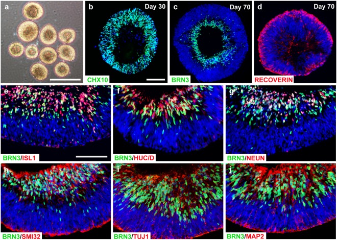
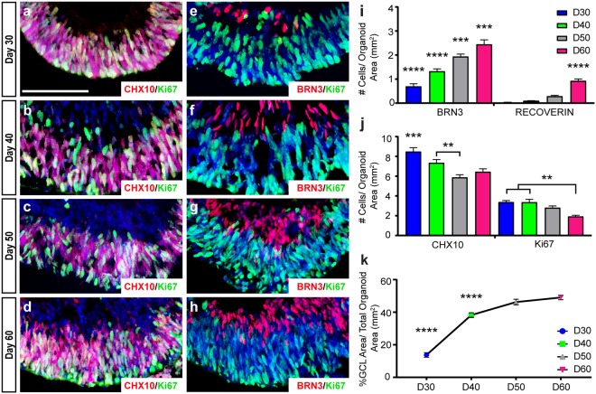
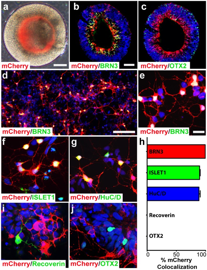
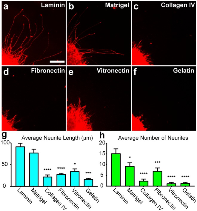
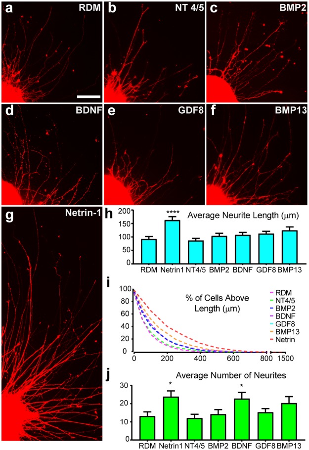
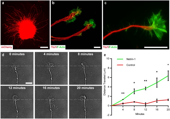
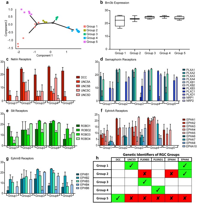
Retinal organoids are three-dimensional structures derived from human pluripotent stem cells (hPSCs) which recapitulate the spatial and temporal differentiation of the retina, serving as effective in vitro models of retinal development. However, a lack of emphasis has been placed upon the development and organization of retinal ganglion cells (RGCs) within retinal organoids. Thus, initial efforts were made to characterize RGC differentiation throughout early stages of organoid development, with a clearly defined RGC layer developing in a temporally-appropriate manner expressing a complement of RGC-associated markers. Beyond studies of RGC development, retinal organoids may also prove useful for cellular replacement in which extensive axonal outgrowth is necessary to reach post-synaptic targets. Organoid-derived RGCs could help to elucidate factors promoting axonal outgrowth, thereby identifying approaches to circumvent a formidable obstacle to RGC replacement. As such, additional efforts demonstrated significant enhancement of neurite outgrowth through modulation of both substrate composition and growth factor signaling. Additionally, organoid-derived RGCs exhibited diverse phenotypes, extending elaborate growth cones and expressing numerous guidance receptors. Collectively, these results establish retinal organoids as a valuable tool for studies of RGC development, and demonstrate the utility of organoid-derived RGCs as an effective platform to study factors influencing neurite outgrowth from organoid-derived RGCs.


Conflict of interest statement

The authors declare no competing interests.

Figures

Figure 1
Figure 1
Organization of a distinct ganglion cell layer within retinal organoids. (a) Brightfield microscopy displayed the stratified morphology of early retinal organoids. (b) Within 30 days of differentiation, nearly all cells within retinal organoids expressed the retinal progenitor marker CHX10. (c–d) Within 70 days of differentiation, a presumptive ganglion cell layer expressing BRN3 occupied basal layers of retinal organoids, while Recoverin-positive photoreceptors resided in more apical layers. (eg) RGCs that were found within the presumptive ganglion cell layer expressed characteristic RGC-associated proteins including BRN3, ISLET1, HuC/D, and NEUN. (hj) BRN3-positive RGCs also co-expressed numerous characteristic cytoskeletal markers such as SMI32, TUJ1, MAP2. Scale bars equal 500μm for (a) and 100μm for (bj). Scale bar in b applies to (c,d), scale bar in e applies to (fj).
Figure 2
Figure 2
Spatial and temporal development of RGCs within retinal organoids. (a–d) The expression of retinal progenitor markers including CHX10 and Ki67 were widely expressed at early stages of organoid development, but became more restricted to outer layers by 60 days of differentiation. (e–h) The restriction of progenitor cells to the outer layers over time was associated with an increase in the expression of the RGC-associated marker BRN3 in the inner layers. (i) Quantification of immunostaining demonstrated a significant increase in BRN3 over time, followed by the delayed onset of expression of photoreceptor markers such as Recoverin. (j) Conversely, the number of CHX10- and Ki67-positive retinal progenitors significantly decreased over time. (k) Correlated with the advancement of RGC differentiation, the presumptive RGC layer area displayed a significant increase in its size over time relative to the size of the organoid. Error bars (n = 3) represent s.e.m. (**p < 0.01, ***p < 0.005, ****p < 0.001). Scale bars equal 100 μm.
Figure 3
Figure 3
Identification of RGCs using a fluorescent reporter. (a–c) RGCs could be readily identified by mCherry expression observed in the inner layers of each organoid, defining the presumptive retinal ganglion cell layer. (d–h) Multiple RGC-associated markers such as BRN3, ISLET1, and HuC/D co-expressed with mCherry. (h–j) Conversely, mCherry did not co-localize with markers of other retinal cells such as Recoverin and OTX2. Error bars represent s.e.m. Scale bars: 100 μm (a–c), 200 μm (d), 20 μm (e–g, i–j).
Figure 4
Figure 4
Substrate Modulation of RGC Neurite Outgrowth. (a–f) mCherry-positive RGCs were analyzed for neurite outgrowth on various substrates including laminin (n = 50), Matrigel (n = 51), collagen IV (n = 28), fibronectin (n = 39), vitronectin (n = 32) and gelatin (n = 16) where n is the number of aggregates analyzed. (g,h) Mean neurite length and mean number of neurites were calculated for each of the experimental conditions. Significant differences from laminin were determined by one-way ANOVA, *p < 0.05, ***p < 0.005, ****p < 0.001. Error bars represent s.e.m. Scale bar: 100 μm.
Figure 5
Figure 5
Modulation of Neurite Outgrowth by Soluble Factors. (a–g) mCherry positive RGCs were analyzed for neurite outgrowth with the addition of various growth factors including NT 4/5 (n = 36), BMP2 (n = 39), BDNF (n = 44), GDF8 (n = 55), BMP13 (n = 44) and Netrin-1 (n = 63) at a concentration of 50 ng/mL, where n is the number of aggregates analyzed. (h) Among the soluble factors tested, Netrin-1 was observed to significantly increase the average length of neurites. (i) All traced neurites were graphed as cumulative frequency above indicated length for each growth factor, with Netrin-1 producing neurites reaching lengths approaching 1.5 mm in 24 hours. (j) The number of neurites was also significantly increased in response to Netrin-1 and BDNF compared to RDM controls. Significance was determined by one-way ANOVA, *p < 0.05, ****p < 0.001. Error bars represent s.e.m. Scale bar: 100 μm.
Figure 6
Figure 6
Identification and Dynamic Rearrangement of RGC Growth Cones (a) Cellular aggregates enriched for mCherry-positive RGCs extended numerous lengthy neurites in all directions after 24 hours in culture. (b) Neurites bundled together and displayed prominent growth cones at the leading edge of neurites. (c) Growth cones exhibited lamellipodia and numerous filopodia enriched for F-actin. (d) DIC time-lapse imaging revealed that these growth cones were also highly dynamic and motile over time, with apparent rearrangement and forward advancement of growth cones. (e) Growth cones exposed to Netrin-1 displayed a significant increase in forward growth compared to untreated controls. Significance was determined with multiple t-tests followed by the Holm-Sidak test. *p < 0.05, **p < 0.01. Error bars represent s.e.m. Scale bars: 150 μm (a), 25 μm (b–d).
Figure 7
Figure 7
Diversity of Guidance Receptor Gene Expression in retinal organoid-derived RGCs. (a) Based on the expression of guidance receptor genes, individual RGCs were analyzed and grouped into five profiles based on gene expression (n = 33). (b) Each of the five groups displayed similar levels of the RGC marker BRN3b. (c–g) Relative gene expression of guidance receptor genes is shown for each of the five groups of RGCs. Gene expression levels are indicated on the Y-axes. Error bars represent s.e.m. (h) Of the 34 guidance receptor genes analyzed, unique expression profiles were generated for each group based on the expression of just 6 genes. Green boxes indicate genes whose expression is required for definitive identification of an RGC group, while red boxes indicate genes whose expression must be absent for definitive identification of that group. Unfilled boxes indicate that the presence or absence of the indicated gene is not a requirement for identification of that particular RGC group.

Similar articles

Cited by

References

    1. Benowitz LI, Yin Y. Optic nerve regeneration. Archives of ophthalmology (Chicago, III.: 1960) 2010;128:1059–1064. doi: 10.1001/archophthalmol.2010.152. - DOI - PMC - PubMed
    1. Crair MC, Mason CA. Reconnecting Eye to Brain. The Journal of neuroscience: the official journal of the Society for Neuroscience. 2016;36:10707–10722. doi: 10.1523/jneurosci.1711-16.2016. - DOI - PMC - PubMed
    1. Laha B, Stafford BK, Huberman AD. Regenerating optic pathways from the eye to the brain. Science (New York, N.Y.) 2017;356:1031–1034. doi: 10.1126/science.aal5060. - DOI - PMC - PubMed
    1. Muramatsu R, Ueno M, Yamashita T. Intrinsic regenerative mechanisms of central nervous system neurons. Bioscience trends. 2009;3:179–183. - PubMed
    1. Shen S, Wiemelt AP, McMorris FA, Barres BA. Retinal ganglion cells lose trophic responsiveness after axotomy. Neuron. 1999;23:285–295. doi: 10.1016/S0896-6273(00)80780-1. - DOI - PubMed

Publication types