

Differentiation enhances Zika virus infection of neuronal brain cells
- PMID: 30266962
- PMCID: PMC6162312
- DOI: 10.1038/s41598-018-32400-7
Differentiation enhances Zika virus infection of neuronal brain cells
Abstract
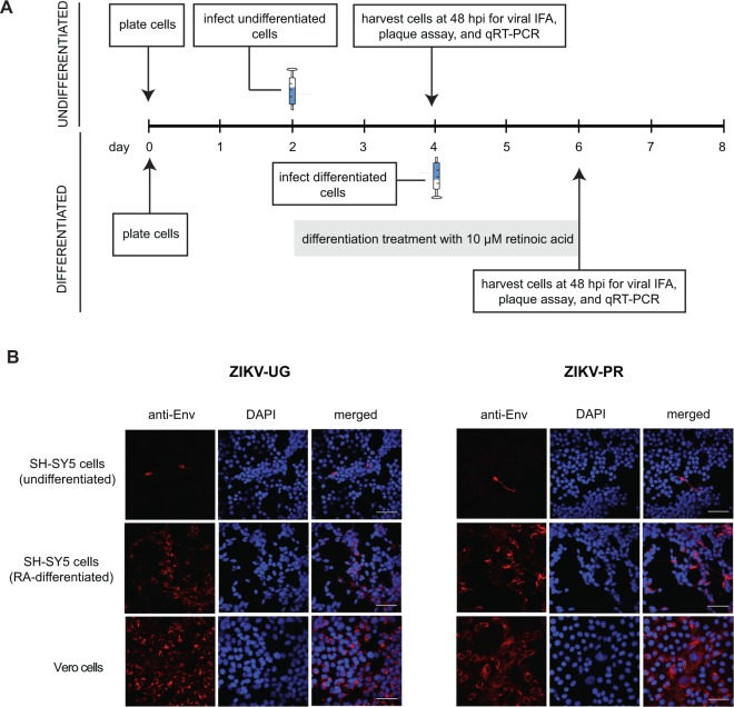
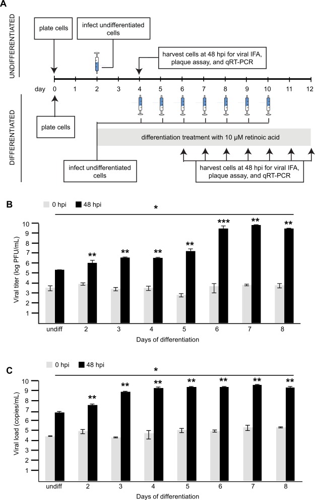
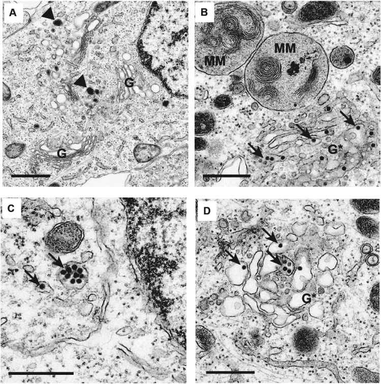
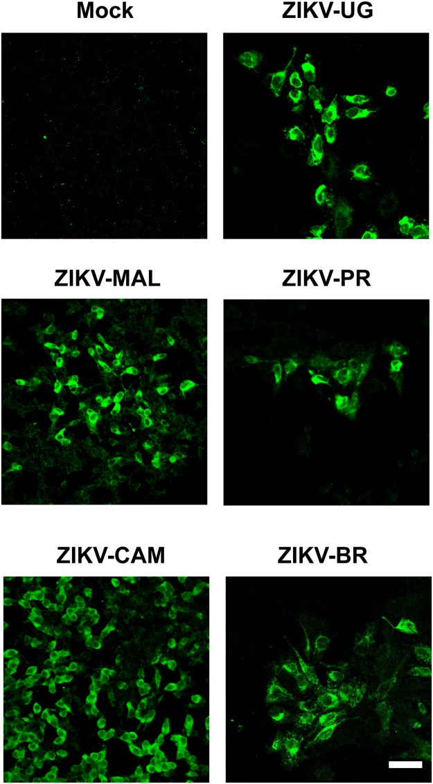
Zika virus (ZIKV) is an emerging, mosquito-borne pathogen associated with a widespread 2015-2016 epidemic in the Western Hemisphere and a proven cause of microcephaly and other fetal brain defects in infants born to infected mothers. ZIKV infections have been also linked to other neurological illnesses in infected adults and children, including Guillain-Barré syndrome (GBS), acute flaccid paralysis (AFP) and meningoencephalitis, but the viral pathophysiology behind those conditions remains poorly understood. Here we investigated ZIKV infectivity in neuroblastoma SH-SY5Y cells, both undifferentiated and following differentiation with retinoic acid. We found that multiple ZIKV strains, representing both the prototype African and contemporary Asian epidemic lineages, were able to replicate in SH-SY5Y cells. Differentiation with resultant expression of mature neuron markers increased infectivity in these cells, and the extent of infectivity correlated with degree of differentiation. New viral particles in infected cells were visualized by electron microscopy and found to be primarily situated inside vesicles; overt damage to the Golgi apparatus was also observed. Enhanced ZIKV infectivity in a neural cell line following differentiation may contribute to viral neuropathogenesis in the developing or mature central nervous system.
Conflict of interest statement
C.Y.C. is the director of the UCSF-Abbott Viral Diagnostics and Discovery Center and receives research support from Abbott Laboratories. The other authors disclose no conflicts of interest.
Figures
Similar articles
-
Infectivity of Immature Neurons to Zika Virus: A Link to Congenital Zika Syndrome.EBioMedicine. 2016 Aug;10:65-70. doi: 10.1016/j.ebiom.2016.06.026. Epub 2016 Jun 23. EBioMedicine. 2016. PMID: 27364784 Free PMC article.
-
Antiviral CD8 T cells induce Zika-virus-associated paralysis in mice.Nat Microbiol. 2018 Feb;3(2):141-147. doi: 10.1038/s41564-017-0060-z. Epub 2017 Nov 20. Nat Microbiol. 2018. PMID: 29158604 Free PMC article.
-
Strain-Dependent Consequences of Zika Virus Infection and Differential Impact on Neural Development.Viruses. 2018 Oct 9;10(10):550. doi: 10.3390/v10100550. Viruses. 2018. PMID: 30304805 Free PMC article.
-
Zika Virus and the Metabolism of Neuronal Cells.Mol Neurobiol. 2019 Apr;56(4):2551-2557. doi: 10.1007/s12035-018-1263-x. Epub 2018 Jul 24. Mol Neurobiol. 2019. PMID: 30043260 Free PMC article. Review.
-
Zika Virus Neuropathogenesis-Research and Understanding.Pathogens. 2024 Jul 2;13(7):555. doi: 10.3390/pathogens13070555. Pathogens. 2024. PMID: 39057782 Free PMC article. Review.
Cited by
-
Zika Virus Impairs Neurogenesis and Synaptogenesis Pathways in Human Neural Stem Cells and Neurons.Front Cell Neurosci. 2019 Mar 15;13:64. doi: 10.3389/fncel.2019.00064. eCollection 2019. Front Cell Neurosci. 2019. PMID: 30949028 Free PMC article.
-
Repression of varicella zoster virus gene expression during quiescent infection in the absence of detectable histone deposition.PLoS Pathog. 2025 Feb 10;21(2):e1012367. doi: 10.1371/journal.ppat.1012367. eCollection 2025 Feb. PLoS Pathog. 2025. PMID: 39928684 Free PMC article.
-
Zika Virus Infects Trabecular Meshwork and Causes Trabeculitis and Glaucomatous Pathology in Mouse Eyes.mSphere. 2019 May 8;4(3):e00173-19. doi: 10.1128/mSphere.00173-19. mSphere. 2019. PMID: 31068433 Free PMC article.
-
The liver X receptor agonist LXR 623 restricts flavivirus replication.Emerg Microbes Infect. 2021 Dec;10(1):1378-1389. doi: 10.1080/22221751.2021.1947749. Emerg Microbes Infect. 2021. PMID: 34162308 Free PMC article.
-
In Vitro Roles of Burkholderia Intracellular Motility A (BimA) in Infection of Human Neuroblastoma Cell Line.Microbiol Spectr. 2023 Aug 17;11(4):e0132023. doi: 10.1128/spectrum.01320-23. Epub 2023 Jul 6. Microbiol Spectr. 2023. PMID: 37409935 Free PMC article.
References
Publication types
MeSH terms
Grants and funding
LinkOut - more resources
Full Text Sources
Other Literature Sources
Medical
