Review
. 2018 Dec;27(12):1984-2009.
doi: 10.1002/pro.3519.

The molecular details of cytokine signaling via the JAK/STAT pathway

Affiliations
Review

The molecular details of cytokine signaling via the JAK/STAT pathway

Rhiannon Morris et al. Protein Sci. 2018 Dec.

Abstract

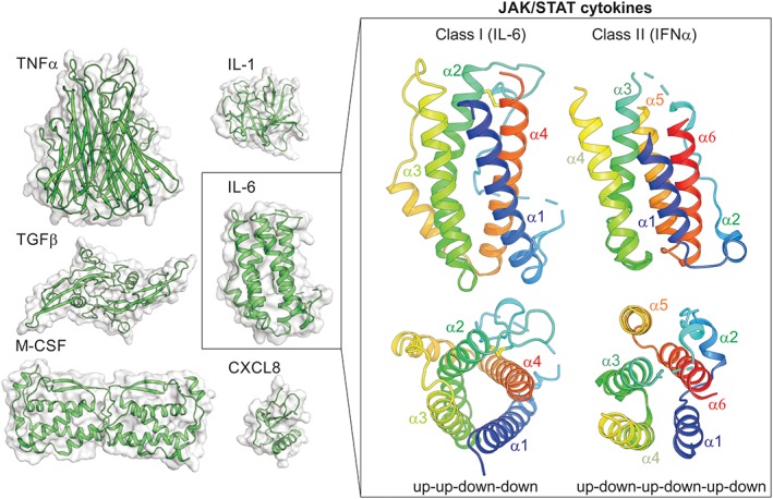
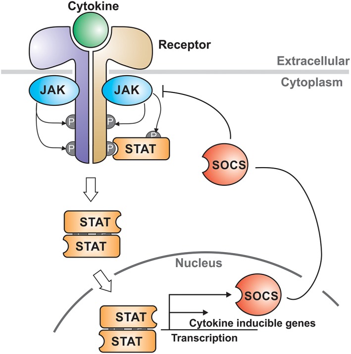
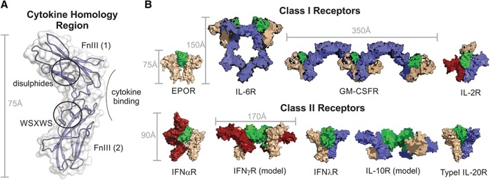
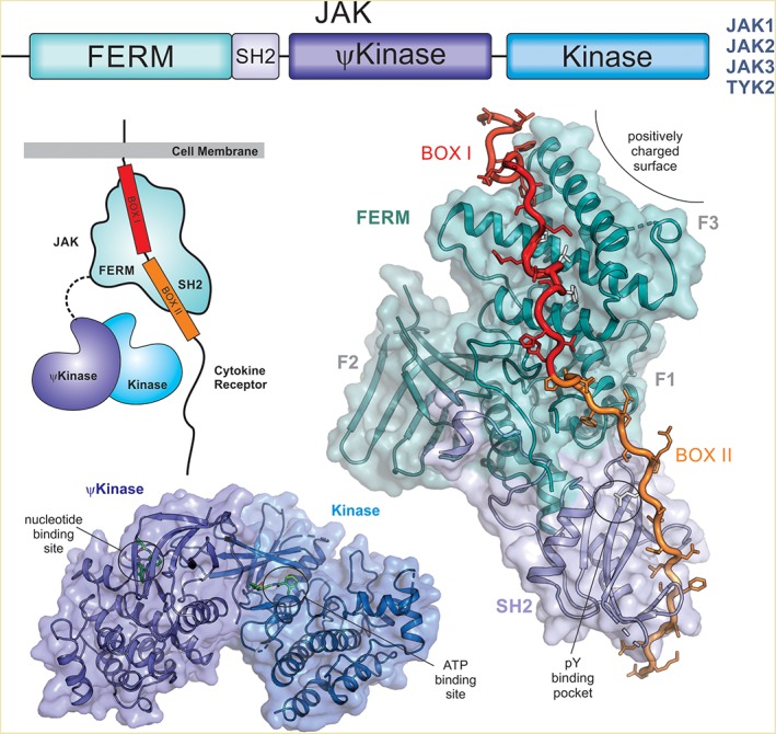
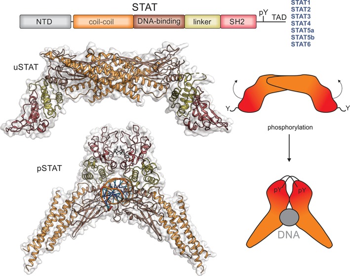
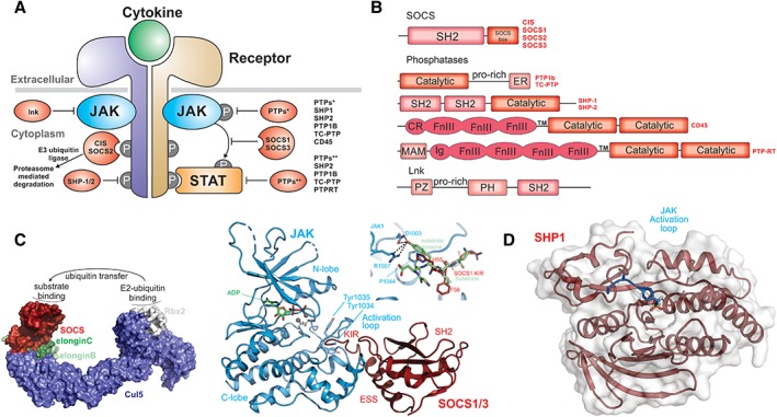
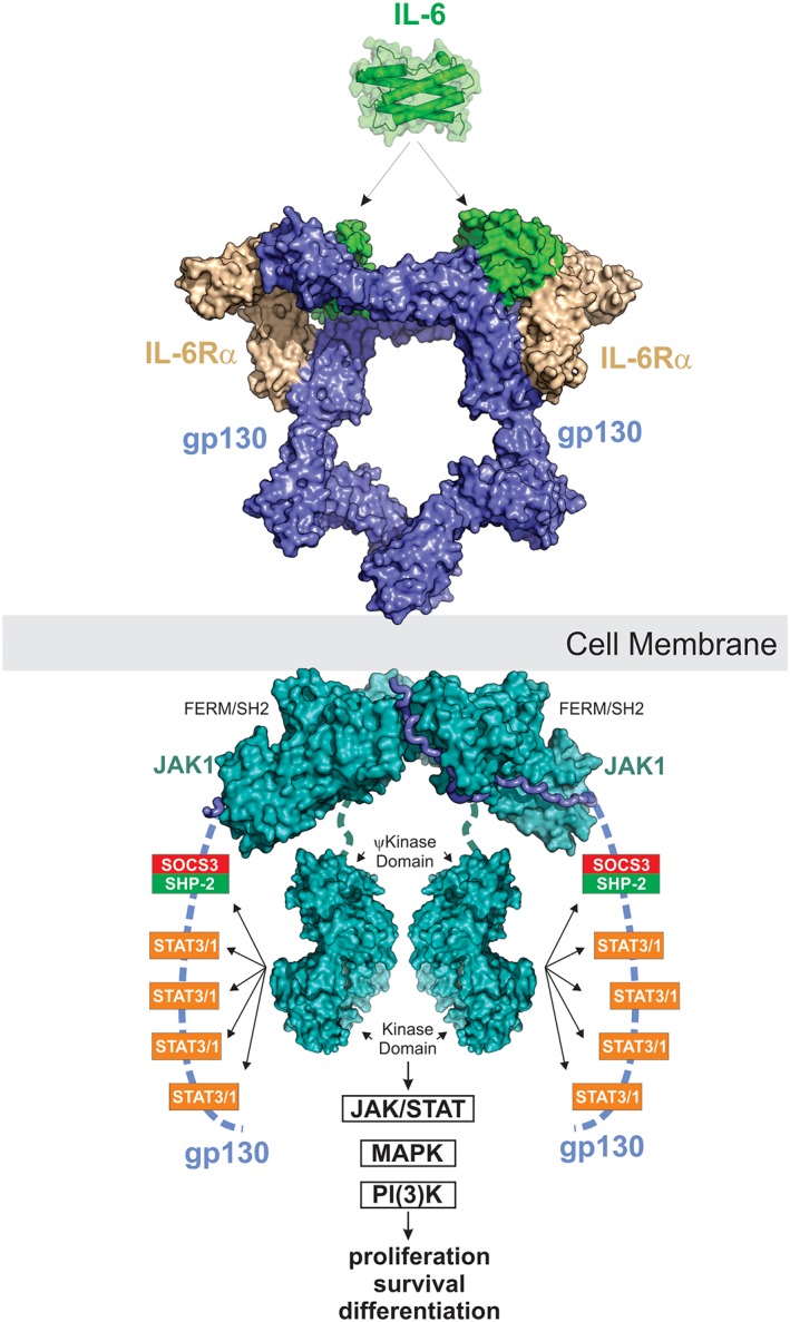
More than 50 cytokines signal via the JAK/STAT pathway to orchestrate hematopoiesis, induce inflammation and control the immune response. Cytokines are secreted glycoproteins that act as intercellular messengers, inducing proliferation, differentiation, growth, or apoptosis of their target cells. They act by binding to specific receptors on the surface of target cells and switching on a phosphotyrosine-based intracellular signaling cascade initiated by kinases then propagated and effected by SH2 domain-containing transcription factors. As cytokine signaling is proliferative and often inflammatory, it is tightly regulated in terms of both amplitude and duration. Here we review molecular details of the cytokine-induced signaling cascade and describe the architectures of the proteins involved, including the receptors, kinases, and transcription factors that initiate and propagate signaling and the regulatory proteins that control it.

Keywords: Cytokine Signaling; JAK/STAT; SOCS; cytokine; cytokine receptor; hematopoiesis.


Figures

Figure 1
Figure 1
Cytokines . Structures of members of the TNFα‐family, TGFβ‐family, IL‐1‐like cytokines, chemokines (CXCL8), cytokines that signal through receptor tyrosine‐kinases (M‐CSF) or the JAK/STAT pathway (IL‐6) are shown on the left. JAK/STAT cytokines are helical bundle cytokines and can be divided into two classes. Examples of these two classes are shown on the right.
Figure 2
Figure 2
The JAK/STAT pathway. Schematic of the signaling cascade induced by cytokines that signal via the JAK/STAT pathway. Cytokine binds to a specific receptor and allows transactivation of the associated Janus Kinases (JAKs). Activated JAKs then phosphorylate tyrosines on the intracellular domains of the receptor which recruit the Signal Transducers and Activators of Transcription (STAT) transcription factors. STATs are translocated into the nucleus and upregulate the transcription of cytokine‐responsive genes. SOCS proteins are direct targets of STAT and act as negative‐feedback inhibitors to switch off the signaling cascade.
Figure 3
Figure 3
Class I and Class II cytokines. Families of cytokines and the receptors they bind to are shown above the JAK‐, STAT‐, and SOCS‐family members they signal through.
Figure 4
Figure 4
The cytokine homology region (CHR) forms the basis of all cytokine receptors. (A) The CHR from a Class I receptor (Growth Hormone Receptor) is shown with the two FnIII domains, disulfide bonds and WSXWS motif highlighted. All receptors contain a CHR however many receptors, especially those that recognize Class I cytokines, have additional FnIII and Ig domains and this results in a large variety of receptor architectures and stoichiometries. (B) Structures and models of a diverse range of cytokine:receptor complexes.
Figure 5
Figure 5
Janus kinases (JAKs). There are four members of the JAK family (JAK1, JAK2, JAK3, and TYK2) and all share similar domain architecture (top). The FERM and SH2 domains tether JAK to the receptor, binding Box I and Box II respectively (structure shown on the right, PDB ID: http://firstglance.jmol.org/fg.htm?mol=5L04)). The pseudokinase (ψkinase) regulates the activity of the catalytically active kinase domain (bottom, PDB ID: http://firstglance.jmol.org/fg.htm?mol=4OLI) via a mechanism that is unclear. There is no structure of a full‐length JAK protein and hence the relative orientation of the N‐ and C‐terminal halves of the protein is unknown (indicated schematically on the left).
Figure 6
Figure 6
STATs. The Signal Tranducers and Activators of Transcription (STATs) are a family of latent transcription factors that are activated by phosphorylation following cytokine exposure. The same domain architecture is shared by all STAT proteins and is shown schematically above. Unphosphorylated STAT (uSTAT) exists as an antiparallel dimer in the cytoplasm (upper). The SH2 domain (red) of uSTAT binds to phosphotyrosines in cytokine receptors which allows JAK to phosphorylate a specific tyrosine located between the SH2 and transactivation domain (TAD). This phosphotyrosine is then targeted by the SH2 domain of the other monomer inducing a large rotation between the two subunits of the dimer and allowing phosphorylated STAT (pSTAT) to occupy its DNA‐binding competent dimeric structure (lower). The structures shown here are of STAT1 (PDB ID: http://firstglance.jmol.org/fg.htm?mol=1YVL,135 http://firstglance.jmol.org/fg.htm?mol=1BF5 136) with the colors matching the schematic representation above. The N‐terminal domain of STAT does not appear to form a stable interaction with the rest of the molecule and is not shown here. The transactivation domain (TAD) is unstructured but allows binding of accessory factors.
Figure 7
Figure 7
Regulation of cytokine signaling. (A) Schematic diagram showing regulators of cytokine signaling and where they act. (B) Domain architecture of the proteins indicated in A. (C) The primary negative feedback regulators of cytokine signaling are a subset of the SOCS (Suppressors of Cytokine Signaling) family, CIS, SOCS1, SOCS2, and SOCS3. These proteins function as the substrate recruitment modules of an E3 ubiquitin ligase (model structure shown in surface representation) and promote the ubiquitination and degradation of cytokine receptors and potentially other substrates. Substrates bind to the SH2 domain of SOCS proteins (red) and ubiquitin is transferred via an E2 ubiquitin‐conjugating enzyme that docks onto the RING‐domain protein Rbx2 (white). SOCS1 and SOCS3 (right) can also directly inhibit the JAK kinase domain by using their kinase inhibitory region (KIR) to block the substrate binding site of the kinase (PDB ID: http://firstglance.jmol.org/fg.htm?mol=6C7Y) (model of a substrate overlay shown inset). (D) Six tyrosine phosphatases have been shown to be important regulators of cytokine‐pathway activity, acting by dephosphorylating JAKs, STATs, or receptors. The structure of one of these, SHP1, has been solved in complex with the JAK activation loop of JAK2 (PDB ID: http://firstglance.jmol.org/fg.htm?mol=4GSO).
Figure 8
Figure 8
IL‐6 signaling. IL‐6 signals via a 2:2:2 complex between itself, gp130 and either membrane‐bound IL‐6Rα (classic signaling) or soluble IL‐6Rα (trans‐signaling). JAK1, JAK2 and TYK2 can all bind the intracellular domain of gp130; however, JAK1 appears to be the dominant kinase. The structure of JAK1 bound to the gp130 cytoplasmic domain is a model based on the structures of JAK1/IFNλR (PDB ID: http://firstglance.jmol.org/fg.htm?mol=5L04) and the JAK2/EPOR dimeric structure (coordinates kindly provided by R. Ferrao and P. Lupardus). JAK is activated by trans‐phosphorylation and then phosphorylates five tyrosine residues on the receptor intracellular domain. The four distal tyrosines are docking sites for STAT3 and to a lesser degree STAT1. Activated STAT3 is then phosphorylated by JAK and translocates into the nucleus to drive the biological response. The MAPK pathway is also stimulated by IL‐6 via SHP2 which binds to pY759 using its SH2 domain. The PI(3)K pathway is also activated in response to IL‐6. SOCS3 is a direct STAT3 target gene and binds to the SHP2‐binding site on the receptor via its SH2 domain. This inhibits MAPK signaling via displacement of SHP2 and also inhibits further STAT3 activation by direct inhibition of JAK catalytic activity.

References

    1. Stark GR, Darnell JE Jr (2012) The JAK‐STAT pathway at twenty. Immunity 36:503–514. - PMC - PubMed
    1. Velazquez L, Fellous M, Stark GR, Pellegrini S (1992) A protein tyrosine kinase in the interferon‐alpha/beta signaling pathway. Cell 70:313–322. - PubMed
    1. Wilks AF (1989) Two putative protein‐tyrosine kinases identified by application of the polymerase chain reaction. Proc Natl Acad Sci U S A 86:1603–1607. - PMC - PubMed
    1. Wilks AF, Harpur AG, Kurban RR, Ralph SJ, Zurcher G, Ziemiecki A (1991) Two novel protein‐tyrosine kinases, each with a second phosphotransferase‐related catalytic domain, define a new class of protein kinase. Mol Cell Biol 11:2057–2065. - PMC - PubMed
    1. Firmbach‐Kraft I, Byers M, Shows T, Dalla‐Favera R, Krolewski JJ (1990) tyk2, prototype of a novel class of non‐receptor tyrosine kinase genes. Oncogene 5:1329–1336. - PubMed

Publication types