. 2018 Nov;7(11):5611-5620.
doi: 10.1002/cam4.1783. Epub 2018 Sep 28.

GNA13 promotes tumor growth and angiogenesis by upregulating CXC chemokines via the NF-κB signaling pathway in colorectal cancer cells

Affiliations

GNA13 promotes tumor growth and angiogenesis by upregulating CXC chemokines via the NF-κB signaling pathway in colorectal cancer cells

Zhongqiang Zhang et al. Cancer Med. 2018 Nov.

Retraction in

  • Retraction.
    [No authors listed] [No authors listed] Cancer Med. 2023 Sep;12(18):19352. doi: 10.1002/cam4.6367. Epub 2023 Sep 19. Cancer Med. 2023. PMID: 37724781 Free PMC article.

Abstract

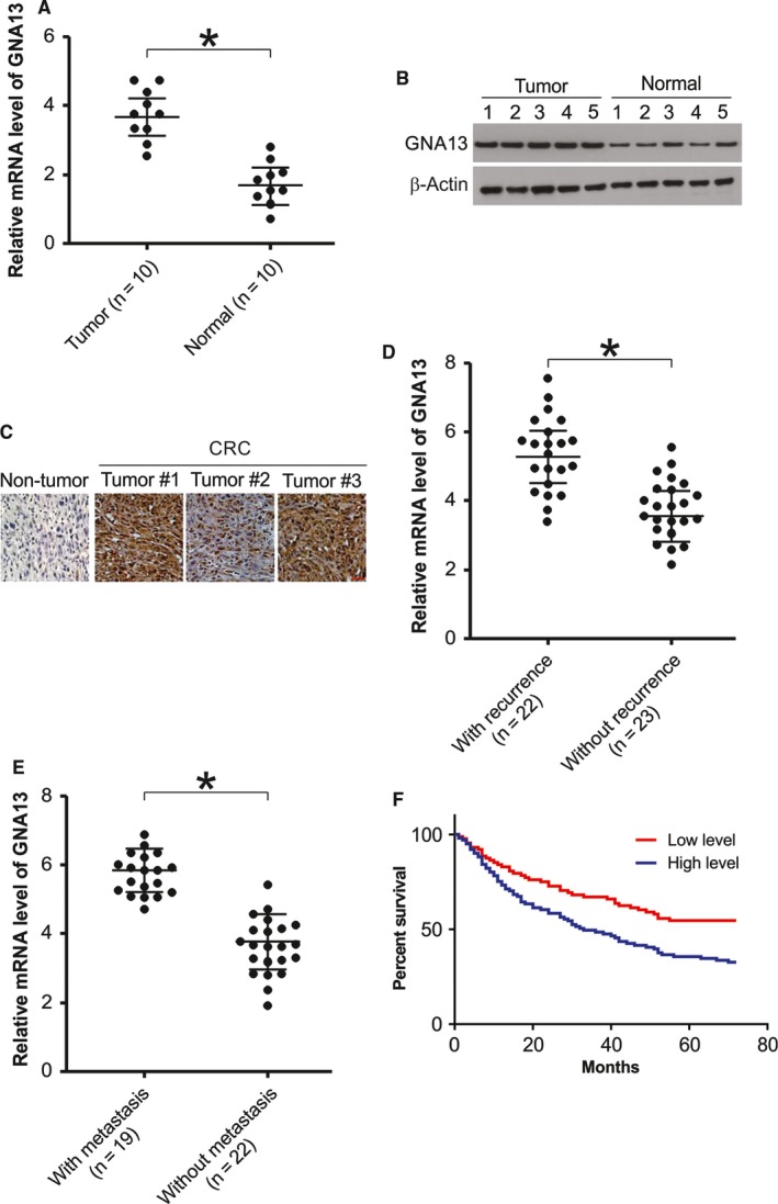
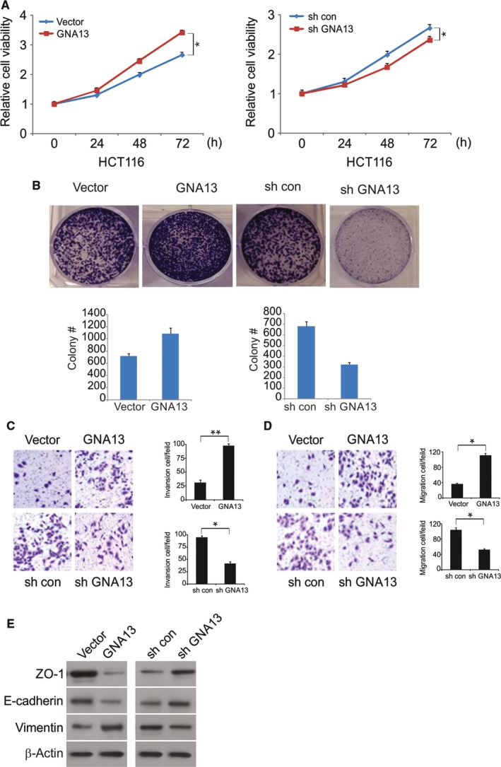
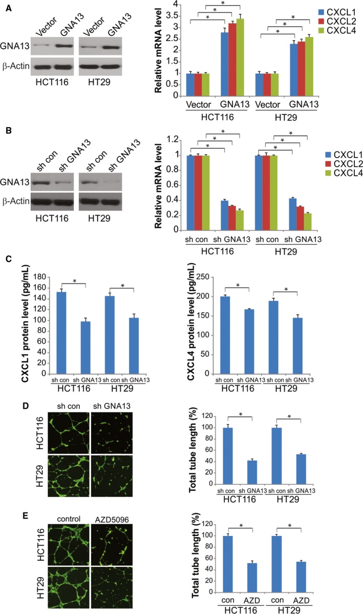
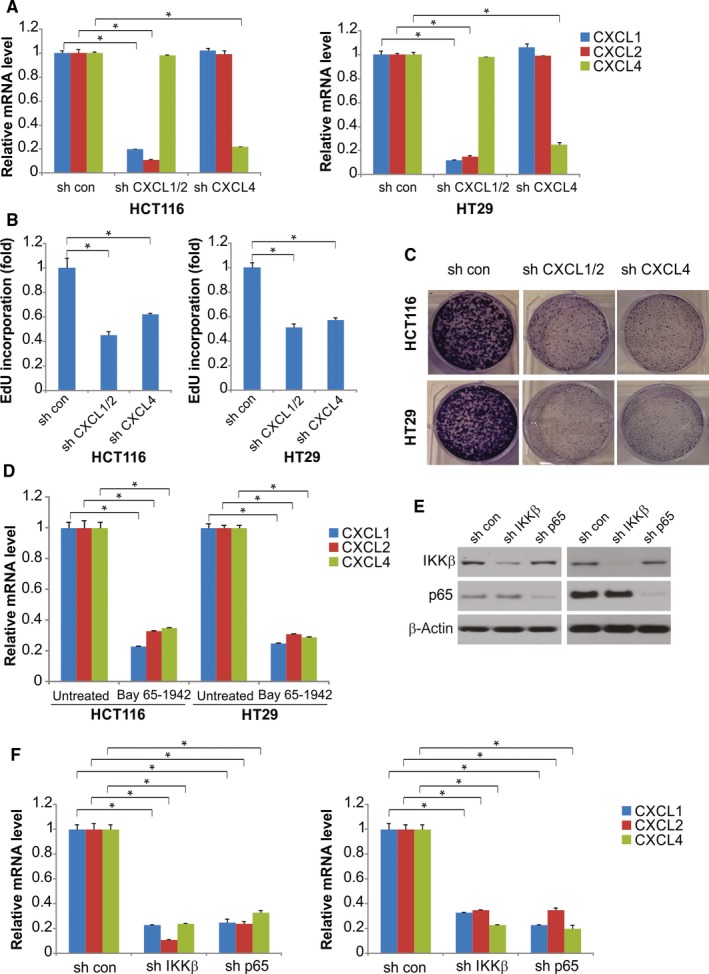
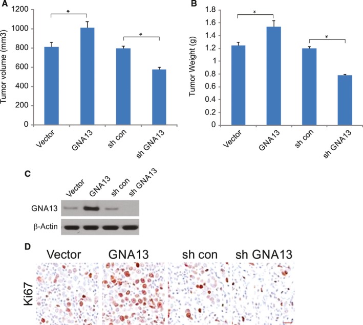
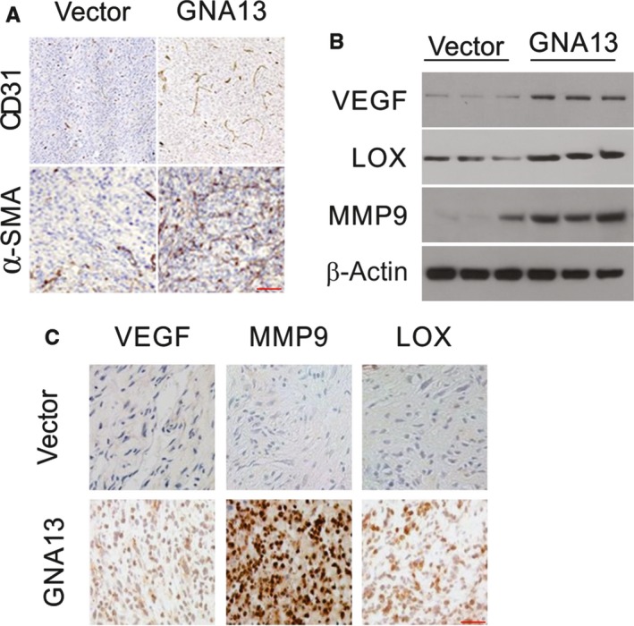
GNA13 has been found overexpressed in various types of cancer, which is related to tumor metastasis and progression. However, the biological functions of GNA13 in colorectal cancer (CRC) progression remain unclear. This study aimed to explore the role of GNA13 in CRC and investigate the mechanism of how GNA13 promotes tumor growth. Interestingly, our findings showed that GNA13 is commonly upregulated in CRC, where these events are associated with a worse histologic grade and poor survival. Increased expression levels of GNA13 promoted cell growth, migration, invasion, and epithelial-mesenchymal transition, whereas GNA13 silencing abrogated these malignant phenotypes. In addition, overexpressing GNA13 in cancer cells increased the levels of the chemokines CXCL1, CXCL2, and CXCL4, which contributed to CRC proliferation and colony formation. Moreover, our mechanistic investigations suggest that the NF-κB/p65 signaling pathway was activated by the increase in GNA13 levels. Inhibiting the NF-κB/p65 pathway with an inhibitor decreased GNA13-induced migration, invasion and CXCL chemokine level increases, indicating the critical role of NF-κB/p65 signaling in mediating the effects of GNA13 in CRC. Together, these results demonstrate a key role of GNA13 overexpression in CRC that contributes to malignant behavior in cancer cells, at least in part through stimulating angiogenesis and increasing the levels of the NF-κB-dependent chemokines CXCL1, CXCL2, and CXCL4.

Keywords: CRC; CXCL chemokines; GNA13; NF-κB.


Figures

Figure 1
Figure 1
GNA13 is overexpressed in CRC and its overexpression in associated with poor prognosis. A, The mRNA level of GNA13 in CRC tissues (Tumor) and paired normal tissues (Normal) was analyzed by real‐time RTPCR. B, GNA13 protein level in CRC tissues and paired normal tissues was assessed by Western blotting. C, Immunohistochemistry of GNA13 in nontumor and primary CRC tissue arrays containing 74 cases. D, Relative mRNA expression of Six1 in HCC samples from patients with disease recurrence (n = 22) or without disease recurrence (n = 23). E, Relative mRNA expression of Six1 in HCC samples from patients with metastasis (n = 19) or without metastasis (n = 22). F, Kaplan‐Meier analysis of overall survival for patients with CRC. The analyses were conducted based on the immunohistochemistry of GNA13 and the survival information provided by the supplier
Figure 2
Figure 2
Effects of GNA13 on malignant phenotypes in CRC cells (A) Cell viability of HCT116 cells was analyzed by MTS assays at different time points. B, Effects of GNA13 on anchorage‐dependent colony formation. (C) GNA13 regulated Transwell cell invasion and (D) Matrigel invasion. E, Expression of epithelial markers, E‐cadherin and ZO‐1, and mesenchymal markers, vimentin, was analyzed by Western blotting. *, < 0.05
Figure 3
Figure 3
Reduced endothelial cell tube formation upon GNA13 suppression and CXCR2 inhibition. A, After 48 h of the transfection of HCT116 and HT29 cells with GNA13 or vector, mRNA expression of CXCL1, CXCL2, and CXCL4 was quantified by qRTPCR (right); GNA13 protein expression was quantified by Western blotting (left). B, Expression of CXCL1, CXCL2, and CXCL4 mRNA in HT29‐GNA13 shRNA, HT29‐control shRNA, HCT116‐GNA13 shRNA, and HCT116‐control shRNA cells was analyzed by qRTPCR. C, Amount of CXCL1 and CXCL4 proteins in cell culture supernatants was determined by ELISA after 24‐hour culture of HCT116 and HT29 cells that stably expressed control shRNA or shRNA against GNA13 in a serum‐free media. D, Left: HUVEC tube formation after HUVECs were incubated on Matrigel for 8 h in the conditioned media from 24‐hour culture of HCT116 and HT29 cells that stably expressed control shRNA or shRNA against GNA13 in a serum‐free media. E, Left: the inhibition of HUVEC tube formation by CXCR2 antagonists. The HUVECs were incubated on Matrigel for 8 h in a mixture of AZD5069 or DMSO with the conditioned media from 24‐hour culture of HCT116 and HT29 cells. *, < 0.05
Figure 4
Figure 4
GNA13 induces CXCL2, CXCL1, and CXCL4 expression through the IKKβ‐NF‐κB pathway. A, HCT116 and HT29 cervical cancer cells were transfected with CXCL1/2, CXCL4, or control shRNA; after 48 h of transfection, expression of CXCL1, CXCL2, and CXCL4 was quantified by qRTPCR. B, In the EdU incorporation assay, 2 d after transfection, reduced expression of CXCL1, CXCL2, or CXCL4 due to shRNAs inhibited HT29 and HCT116 cell proliferation compared with that in the cells transfected with control shRNA. C, Suppression of CXCL1, CXCL2, or CXCL4 by shRNAs inhibits the clonogenic growth of HCT116 and HT29 cells. D, Expression of CXCL1, CXCL2, and CXCL4 mRNA in HCT116 and HT29 cells treated with 2 μm IKKβ inhibitor Bay 65‐1942 or the control was analyzed by qRTPCR. E, IKKβ and NF‐κB p65 protein expression was quantified by Western blotting. F, HT29 and HCT116 cells were transfected with shRNA against IKKβ, NF‐κB p65, or the control, after 48 h of transfection, and then expression of CXCL1, CXCL2, and CXCL4 mRNA was quantified by qRTPCR. *, < 0.05
Figure 5
Figure 5
GNA13 enhanced tumor growth in vivo (A) Tumor volume in each group. B, Tumor weight in each group. C, The expression of GNA13 in each group was detected by Western blotting. D, Expression of Ki‐67 (cell proliferation marker) in xenograft tumors analyzed by immunohistochemistry. *, < 0.05
Figure 6
Figure 6
GNA13 overexpression promotes angiogenesis (A) Immunofluorescent staining of CD31 (red) and α‐SMA (red) merged with nuclei (blue) in vector and GNA13 HCT116 tumors. B, VEGF, MMP9, MMP2, and LOX in sera from GNA13 tumor‐bearing mice and vector mice were analyzed by Western blotting. C, The expression of MMP9, VEGF, and LOX in GNA13 and vector tumors as assessed by immunohistochemistry

References

    1. De Rosa M, Pace U, Rega D, et al. Genetics, diagnosis and management of colorectal cancer (Review). Oncol Rep. 2015;34:1087‐1096. - PMC - PubMed
    1. Martini G, Troiani T, Cardone C, et al. Present and future of metastatic colorectal cancer treatment: a review of new candidate targets. World J Gastroenterol. 2017;23:4675‐4688. - PMC - PubMed
    1. Peluso G, Incollingo P, Calogero A, et al. Current tissue molecular markers in colorectal cancer: a literature review. Biomed Res Int. 2017;2017:2605628. - PMC - PubMed
    1. Wu F, Song G, de Graaf C, Stevens RC. Structure and function of peptide‐binding G protein‐coupled receptors. J Mol Biol. 2017;429:2726‐2745. - PubMed
    1. Latek D, Bajda M, Filipek S. A hybrid approach to structure and function modeling of G protein‐coupled receptors. J Chem Inf Model. 2016;56:630‐641. - PubMed

Publication types