. 2019 Jan;12(1):36-42.
doi: 10.1016/j.tranon.2018.09.004. Epub 2018 Sep 26.

Smoking-Induced SLPI Expression Hinders HPV Infections Also in Squamous Cell Carcinomas of the Vulva

Affiliations

Smoking-Induced SLPI Expression Hinders HPV Infections Also in Squamous Cell Carcinomas of the Vulva

Elgar S Quabius et al. Transl Oncol. 2019 Jan.

Abstract

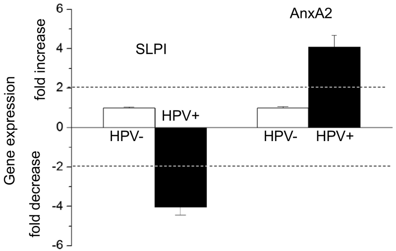
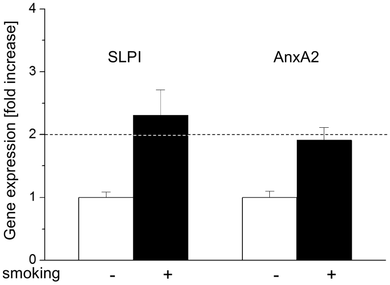
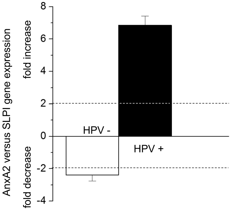
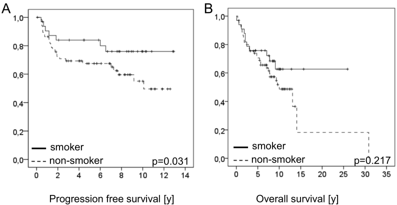
In HNSCC, protein- and mRNA-expression of the antileukoproteinase SLPI are significantly inverse correlated with HPV-infection suggesting that elevated expression of SLPI protects against HPV-infections. Moreover, SLPI-expression is up-regulated in HNSCC-patients reporting a smoking habit. Here, we investigate the described correlation in other HPV-driven cancers, namely vulvar squamous cell carcinoma (VSCC). FFPE samples of 99 VSCC were analyzed by PCR for HPV-DNA-expression and by RT-qPCR for SLPI-mRNA-expression. Of 99 VSCC 10 (10.1%) are HPV-positive; 9 were HPV16; 1 HPV18; all were E6/E7 mRNA-positive. 33 of the 99 patients (33.3%) reported a smoking habit; 7 (21.1%) of these were HPV-positive. Of 66 (66.7%) non-smokers 3 (4.5%) were HPV-positive. SLPI-expression was 4.0-fold lower in HPV-positive than HPV-negative patients. Smoking resulted in 2.3-fold higher SLPI expression. The data presented here indicate that SLPI plays a pivotal role in HPV-infection not only in HNSCC but also in VSCC and possibly also in other HPV-driven cancers. This however, needs to be analyzed in future studies. Furthermore these data lead to the hypothesis that the smoking induced SLPI-increase is systemic rather than local, as assumed based on the HNSCC data.


Figures

Figure 1
Figure 1
Effect of HPV-status on SLPI and AnxA2 gene expression. SLPI and AnxA2 gene expression in all 99 VSCC patients is shown. ΔΔct values obtained in HPV negative patients were set as “1” and fold changes of HPV-positive patients were calculated as described elsewhere (Pfaffl MW: A new mathematical model for relative quantification in real-time RT-PCR. Nucleic Acids Res 29: e45, 2001). Dotted lines indicate 2-fold changes of gene expression (both decrease and increase) indicating, as described previously (Pfaffl MW: A new mathematical model for relative quantification in real-time RT-PCR. Nucleic Acids Res 29: e45, 2001), significant changes in gene expression levels.
Figure 2
Figure 2
Effect of smoking on SLPI and AnxA2 gene expression. SLPI and AnxA2 gene expression is shown. The ΔΔct values obtained in non-smoking patients were set as “1” and fold changes of smoking patients were calculated as described elsewhere (Pfaffl MW: A new mathematical model for relative quantification in real-time RT-PCR. Nucleic Acids Res 29: e45, 2001). Dotted lines indicate 2-fold changes of gene expression indicating, as described previously (Pfaffl MW: A new mathematical model for relative quantification in real-time RT-PCR. Nucleic Acids Res 29: e45, 2001), significant changes in gene expression levels.
Figure 3
Figure 3
Comparison of AnxA2 and SLPI gene expression. The comparison of AnxA2 and SLPI gene expression is shown. To compare AnxA2 versus SLPI gene expression levels, SLPI gene expression for each patient group was set as “1”. Fold change expression levels of AnxA2 were then calculated as described elsewhere (Pfaffl MW: A new mathematical model for relative quantification in real-time RT-PCR. Nucleic Acids Res 29: e45, 2001). Dotted lines indicate 2-fold changes of gene expression level (both decrease and increase) as described previously (Pfaffl MW: A new mathematical model for relative quantification in real-time RT-PCR. Nucleic Acids Res 29: e45, 2001); ± 2-fold changes in gene expression levels are considered statistically significant.
Figure 4
Figure 4
Overall and progression free survival of patients with VSCC in relation to HPV status and SLPI expression. In Figure 4A and B, PFS and in Figure 4C and D OS is shown. PFS for HPV-negative patients (n = 89) was 72.4%, 71.1 and 59.3 after 3, 5, and 10 years respectively. HPV-positive patients (n = 10) showed 88.9% survival after 3 and 5 years. OS of the HPV-negative patients (n = 89) was 78.4%, 71.1% and 55.8% after 3, 5, and 10 years respectively and for the HPV-positive patients (n = 10) OS was 80% both after 3 and 5 years. For Kaplan–Meier analysis SPLI expression of tumors with negative and weak expression (n = 56) and for tumors with moderate and strong expression (n = 43) was pooled. Patients with negative/weak expression showed 78.0% PSF after 3 and 5 years and of 68.9% after 10 years, while patients with moderate/strong SLPI tumor expression showed 69.0% after 3 years, 66.5% after 5 years and 52.5% PFS after 10 years (P = .044). OS of patients with negative/ weak and moderate/strong SLPI expression was not significantly different (P = .502), with OS in patients with negative/ weak SLPI expression being 78.6% after 3 years, 71.2% after 5 years and 52.0% after 10 years. OS of the patients with moderate/strong expression was 78.7%, 73.7% and 60.2% after 3, 5, and 10 years respectively.
Figure 5
Figure 5
Overall and progression free survival of patients with VSCC in relation to smoking habit of the patients. In Figure 5A PFS and in Figures 5B OS is shown. PFS of the patients reporting to be non-smokers (n = 66) was 67.7%, 65.9% and 53.28% after 3, 5, and 10 years respectively, while PFS of patients reporting a smoking habit was 87.1% after 3 and 5 years and 79.2% 10 years (P = .031). OS in non-smoking patients was 78.5%, 70.5% and 51.7% after 3, 5, and 10 years respectively, while in smoking patients 3, 5 and 10 year OS survival rates were 78.8%, 75.(% and 62.7% respectively (P = 0.217).

References

    1. Faraji F, Zaidi M, Fakhry C, Gaykalova DA. Molecular mechanisms of human papillomavirus-related carcinogenesis in head and neck cancer. Microbes Infect. 2017;19:464–475. - PMC - PubMed
    1. Quabius ES, Haag J, Kuhnel A, Henry H, Hoffmann AS, Gorogh T, Hedderich J, Evert M, Beule AG, Maune S. Geographical and anatomical influences on human papillomavirus prevalence diversity in head and neck squamous cell carcinoma in Germany. Int J Oncol. 2015;46:414–422. - PubMed
    1. Hoffmann M, Ihloff AS, Gorogh T, Weise JB, Fazel A, Krams M, Rittgen W, Schwarz E, Kahn T. p16(INK4a) overexpression predicts translational active human papillomavirus infection in tonsillar cancer. Int J Cancer. 2010;127:1595–1602. - PubMed
    1. Klussmann JP, Weissenborn SJ, Wieland U, Dries V, Eckel HE, Pfister HJ, Fuchs PG. Human papillomavirus-positive tonsillar carcinomas: a different tumor entity? Med Microbiol Immunol. 2003;192:129–132. - PubMed
    1. Prigge ES, von Knebel DM, Reuschenbach M. Clinical relevance and implications of HPV-induced neoplasia in different anatomical locations. Mutat Res Rev Mutat Res. 2017;772:51–66. - PubMed

LinkOut - more resources