. 2018 Dec:65:29-36.
doi: 10.1016/j.intimp.2018.09.013. Epub 2018 Sep 27.

Antimicrobial and immunomodulatory activity induced by loperamide in mycobacterial infections

Affiliations

Antimicrobial and immunomodulatory activity induced by loperamide in mycobacterial infections

Esmeralda Juárez et al. Int Immunopharmacol. 2018 Dec.

Abstract

  1. Loperamide modulates macrophages immune responses towards mycobacteria.

  2. Loperamide is an immunoregulator of inflammation during mycobacterial infection.

  3. Loperamide induces immunomodulatory responses and bactericidal mechanisms.

  4. The activation of opioid receptors by loperamide is involved in its immunomodulatory activity.

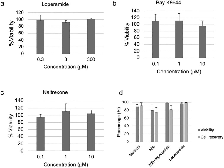
Loperamide is an antidiarrheal drug that targets μ-opioid receptors and calcium channels. A previous report demonstrated that loperamide induces autophagy and enhances antimicrobial activity towards M. tuberculosis in murine and human alveolar macrophages. The aim of this study was to evaluate the immunomodulatory effects of loperamide on macrophages with respect to cytokine and antimicrobial peptide production during mycobacterial infection. We infected monocyte-derived macrophages (macrophages) with M. tuberculosis H37Rv at a multiplicity of infection (MOI) of 5 and treated the cells with 3 μM loperamide. Cytokine production in the supernatants of 24-h cultures and gene expression of the cytokines TNFα, IL1β and IL10 and the antimicrobial peptides LL37 and bactericidal/permeability increasing protein (BPI) in the cell lysates was measured. Intracellular bacterial loads were evaluated by enumerating colony-forming units 3 days posttreatment for M. tuberculosis and 24 h posttreatment for M. smegmatis. We observed that loperamide exerted an immunomodulatory effect on TNFα production in human macrophages infected with M. tuberculosis and that these responses were independent of the bacteria, as they also occurred when macrophages were infected with M. smegmatis and to a lesser extent with M. bovis. In addition, antibacterial mechanisms triggered by loperamide induced a significant reduction in bacterial load and an upregulation of BPI and LL37 gene expression. Thus, our results show that loperamide exerts immunomodulatory effects, which supports its use for additional medical conditions other than diarrhea.

Keywords: BPI; Immunomodulatory activity; LL37; Loperamide; Opioid receptors; Tuberculosis.


Figures

Fig. 1
Fig. 1
Loperamide induces antimicrobial responses in human macrophages infected with M. tuberculosis. Macrophages were infected with M. tuberculosis H37Rv at an MOI of 5 for 1 h. After extensive washing, the cells were treated with 3 μM loperamide for an additional 3 days (a) or 24 h (b and c). The intracellular bacterial loads were evaluated by counting the CFUs (a) (n = 9, *p < 0.05). Cell lysates were obtained for quantitative PCR to assess BPI protein (n = 13) and cathelicidin LL37 (n = 11) levels relative to those of unstimulated cells, with experiments performed in duplicate. The individual values are depicted. *p < 0.05 vs control.
Fig. 2
Fig. 2
Loperamide modulates the proinflammatory environment of infected macrophages. Macrophages were infected with M. tuberculosis H37Rv at an MOI of 5 for 1 h. After extensive washing, the cells were treated with 3 μM loperamide for an additional 24 h. The production of IL1β, IL10, and TNFα was measured in the culture supernatants using a customized Luminex assay, and PGE2 was measured by ELISA (left). The individual values are depicted; *p < 0.05, Friedman's ANOVA followed by Dunn's multiple comparison test. Cell lysates were obtained to assess IL1β, IL10, TNFα and COX-2 gene expression in infected cells treated with loperamide relative to that in untreated infected cells by quantitative PCR, with experiments performed in duplicate. The individual values are depicted. *p < 0.05 vs control.
Fig. 3
Fig. 3
Loperamide downregulates TNFα production in mycobacterium-infected macrophages. (a) Cells were stimulated with 3 μM loperamide or 100 ng/mL LPS for 24 h. TNFα (n = 10) and IL10 (n = 14) production was determined in the culture supernatants by ELISA. The individual results are depicted, and lines indicate the medians; *p < 0.05, ns = not significant. (b) Cells were infected with M. tuberculosis H37Rv at an MOI of 5 for 1 h. After extensive washing, the cells were treated with 3 μM loperamide for an additional 3 days (left) or 24 h (right). The intracellular bacterial loads were evaluated by counting the CFUs, and the levels of TNFα in the culture supernatants were determined by ELISA. CFUs are depicted as medians with interquartile range and TNFα as boxplots indicating quartiles and medians, n = 6, *p < 0.05 vs medium. EtOH, ethyl alcohol, used as control. (c) Cells were treated with 100 ng mL−1 LPS or infected with M. bovis BGC or M. smegmatis at an MOI of 10 for 1 h. After extensive washing to discard nonphagocytized bacteria, the cells were treated with 3 μM loperamide for additional 24 h. TNFα production was determined in the culture supernatants by ELISA. Box plots indicate quartiles and medians, n = 11. °p < 0.05 vs medium, *p < 0.05 between selected pairs, ns = not significant, Friedman's ANOVA followed by Dunn's multiple comparison test.
Fig. 4
Fig. 4
Effect of increased intracellular calcium influx or opioid receptor blockade on immunomodulation by loperamide. Macrophages were infected with M. smegmatis at an MOI of 10 for 1 h. After extensive washing to discard nonphagocytized bacteria, cells were treated with increasing concentrations of (a) Bay K8647 or (b) naltrexone for 30 min followed by treatment with 3 μM loperamide for an additional 24 h. TNFα production was in the culture supernatants was assessed by ELISA. Box plots indicate quartiles and medians, n = 10. °p < 0.05 vs medium, *p < 0.05 between selected pairs, Friedman's ANOVA followed by Dunn's multiple comparison test.
Fig. 5
Fig. 5
Effect of increased intracellular calcium influx or opioid receptor blockade on the antimicrobial activity induced by loperamide. Macrophages were infected with M. smegmatis at an MOI of 10 for 1 h. After extensive washing to remove nonphagocytized bacteria, the cells were treated with 1 μM Bay K8647 or naltrexone for 30 min followed by treatment with 3 μM loperamide for an additional 24 h. The intracellular bacterial loads were evaluated by counting the CFUs (a); n = 8, *p < 0.05 between selected pairs. TNFα production was determined in the culture supernatants by ELISA (b). EtOH and DMSO were used as controls. Box plots indicate quartiles and medians, n = 8, *p < 0.05 vs medium, Friedman's ANOVA followed by Dunn's multiple comparison test.
Fig. 6
Fig. 6
Loperamide-dependent BPI and LL37 gene expression are regulated by μ-opioid receptor signaling. Macrophages were infected with M. smegmatis (a, b) or M. tuberculosis (c, d) at an MOI of 5 for 1 h. After extensive washing to discard nonphagocytized bacteria, the cells were treated with 1 μM naltrexone for 30 min followed by treatment with 3 μM loperamide for an additional 24 h. Cell lysates were obtained to assess BPI and LL37 gene expression relative to that of untreated mycobacterium-infected cells by quantitative PCR, with experiments performed in duplicate. The means ± SEM of 6 independent experiments are depicted. *p < 0.05 vs control.
Supplementary Fig. 1
Cell viability of the chemicals used in this study. Macrophages were stimulated with increasing concentrations of Loperamide (a), Bay K8644 (b) and Naltrexone (c) for 24 h. The cell viability was measured using Cell Titer (Promega) following the manufacturers' instructions. The percentage of viability was calculated relative to unstimulated cells. The Mean ± SE of 5 independent experiments are depicted. (d) Macrophages were cultured during 3 days in presence or absence of infection and loperamide. The cells were detached and counted to determine the number of cells and the viability was calculated by trypan blue exclusion. The Mean percentages ± SE of 3 independent experiments are depicted.
Supplementary Fig. 2
Loperamide induce the production of significant amounts of IL6 (n = 15) and of GM-CSF (n = 8) in macrophages infected with M. tuberculosis, but not of IL12p70 and IL8. The macrophages were infected with M. tuberculosis H37 Rv at an MOI of 5 for 1 h. After extensive washing, the cells were treated with loperamide 3 μM or 100 ng ml−1 of LPS for additional 24 h. The cytokines were measured in the supernatants by luminex. *p < 0.05, vs medium.

References

    1. Juárez E., Carranza C., Sánchez G., González M., Chávez J., Sarabia C., Torres M., Sada E. Loperamide restricts intracellular growth of mycobacterium tuberculosis in lung macrophages. Am. J. Respir. Cell Mol. Biol. 2016 doi: 10.1165/rcmb.2015-0383OC. rcmb.2015-0383OC. - DOI - PubMed
    1. Baker D.E. Loperamide: a pharmacological review. Rev. Gastroenterol. Disord. 2007;7(Suppl. 3):S11–S18. - PubMed
    1. Corsetti M., Whorwell P. Novel pharmacological therapies for irritable bowel syndrome. Expert Rev. Gastroenterol. Hepatol. 2016;10:807–815. doi: 10.1586/17474124.2016.1158099. - DOI - PubMed
    1. Hagiwara K., Nakagawasai O., Murata A., Yamadera F., Miyoshi I., Tan-No K., Tadano T., Yanagisawa T., Iijima T., Murakami M. Analgesic action of loperamide, an opioid agonist, and its blocking action on voltage-dependent Ca2+ channels. Neurosci. Res. 2003;46:493–497. - PubMed
    1. He L.-P., Mears D., Atwater I., Rojas E., Cleemann L. Loperamide mobilizes intracellular Ca2+ stores in insulin-secreting HIT-T15 cells. Br. J. Pharmacol. 2003;139:351–361. doi: 10.1038/sj.bjp.0705263. - DOI - PMC - PubMed

MeSH terms