Review
. 2018 Sep 28:2018:bcr2018225625.
doi: 10.1136/bcr-2018-225625.

Perianeurysmal vasogenic oedema (PAVO) following aneurysm embolisation: a unique case of asymptomatic long-term progression and review of the literature

Affiliations
Review

Perianeurysmal vasogenic oedema (PAVO) following aneurysm embolisation: a unique case of asymptomatic long-term progression and review of the literature

Anthony Ernest Robinson et al. BMJ Case Rep. .

Abstract

Perianeurysmal vasogenic oedema is a recognised although rare phenomenon following endovascular treatment of certain intracranial aneurysms. We present a unique case of asymptomatic perianeurysmal vasogenic oedema following bare platinum coil embolisation of an incidentally discovered right middle cerebral artery aneurysm that slowly increased over a period of 6 years before stabilising and regressing. During this time, the coiled aneurysm per se remained completely stable on serial magnetic resonance angiography.

Keywords: interventional radiology; neuroimaging.


Conflict of interest statement

Competing interests: None declared.

Figures
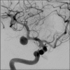
Figure 1
Figure 1
Digital subtraction angiography demonstrated narrow-necked middle cerebral artery (M1 segment) aneurysm measuring 9.7 mm in diameter.
Figure 2
Figure 2
Postcoiling angiography demonstrated residual minor body filling. However, there was persistent intra-aneurysmal stasis of contrast (despite full systemic heparinisation and dual antiplatelet cover) from (A) early arterial, (B) early venous through to (C) late venous phase indicative of impending thrombosis.
Figure 3
Figure 3
T2-weighted axial MRI images 3 years post-treatment (A1–A5) and corresponding images 6 years post-treatment (B1–B5) demonstrating progressive perianeurysmal vasogenic oedema.

References

    1. Szikora I, Seifert P, Hanzely Z, et al. . Histopathologic evaluation of aneurysms treated with Guglielmi detachable coils or matrix detachable microcoils. AJNR Am J Neuroradiol 2006;27:283–8. - PMC - PubMed
    1. Hayashi K, Kitagawa N, Morikawa M, et al. . Long-term follow-up of endovascular coil embolization for cerebral aneurysms using three-dimensional time-of-flight magnetic resonance angiography. Neurol Res 2009;31:674–80. 10.1179/174313209X380964 - DOI - PubMed
    1. Horie N, Kitagawa N, Morikawa M, et al. . Progressive perianeurysmal edema induced after endovascular coil embolization. Report of three cases and review of the literature. J Neurosurg 2007;106:916–20. 10.3171/jns.2007.106.5.916 - DOI - PubMed
    1. Su IC, Willinsky RA, Fanning NF, et al. . Aneurysmal wall enhancement and perianeurysmal edema after endovascular treatment of unruptured cerebral aneurysms. Neuroradiology 2014;56:487–95. 10.1007/s00234-014-1355-x - DOI - PubMed
    1. Lukic S, Jankovic S, Popovic KS, et al. . Analysis of risk factors for perifocal oedema after endovascular embolization of unruptured intracranial arterial aneurysms. Radiol Oncol 2015;49:341–6. 10.1515/raon-2015-0044 - DOI - PMC - PubMed