. 2018 Nov;247(11):1227-1236.
doi: 10.1002/dvdy.24675. Epub 2018 Nov 7.

Comparative Analysis of cul5 and rbx2 Expression in the Developing and Adult Murine Brain and Their Essentiality During Mouse Embryogenesis

Affiliations

Comparative Analysis of cul5 and rbx2 Expression in the Developing and Adult Murine Brain and Their Essentiality During Mouse Embryogenesis

Keiko Hino et al. Dev Dyn. 2018 Nov.

Abstract

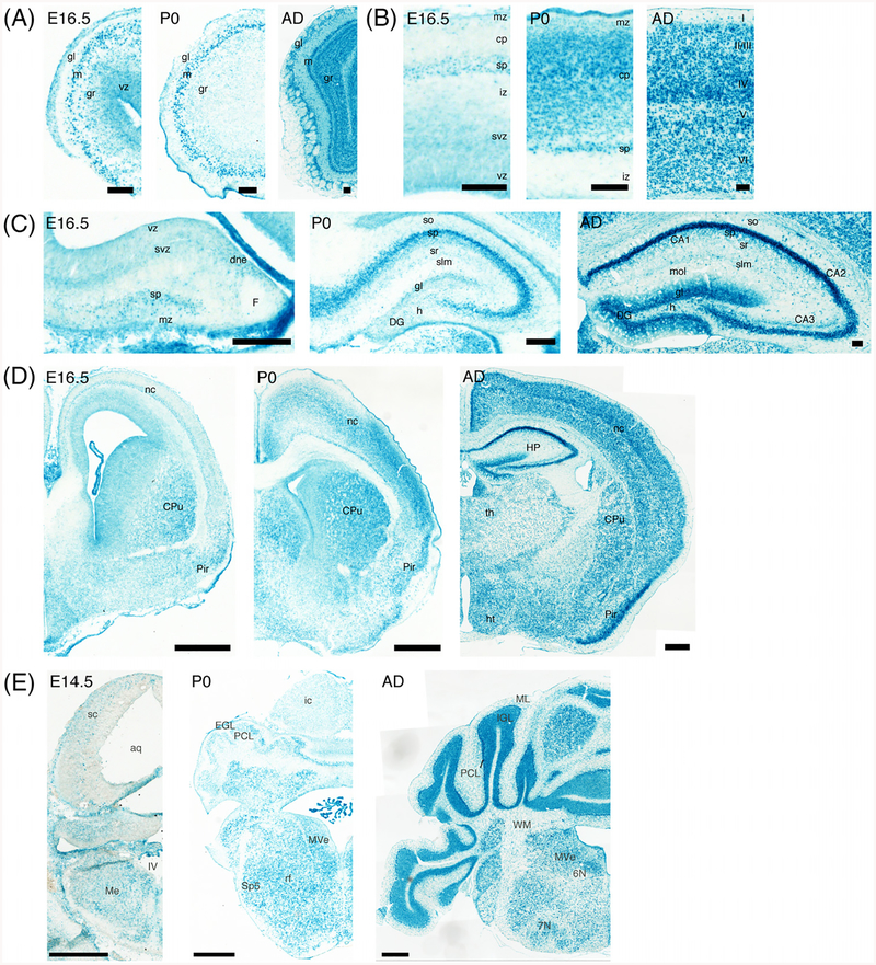
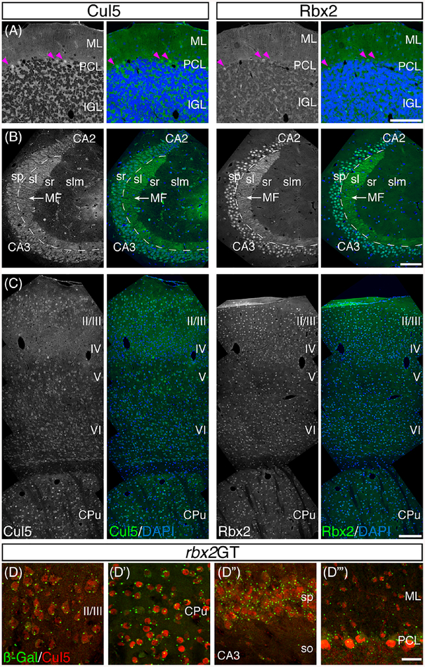
Background: The E3 Cullin 5-RING ubiquitin ligase (CRL5) is a multiprotein complex that has recently been highlighted as a major regulator of central nervous system development. Cullin 5 (Cul5) and the RING finger protein Rbx2 are two CRL5 core components required for CRL5 function in the brain, but their full expression patterns and developmental functions have not been described in detail.

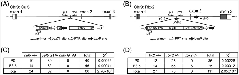
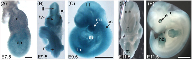
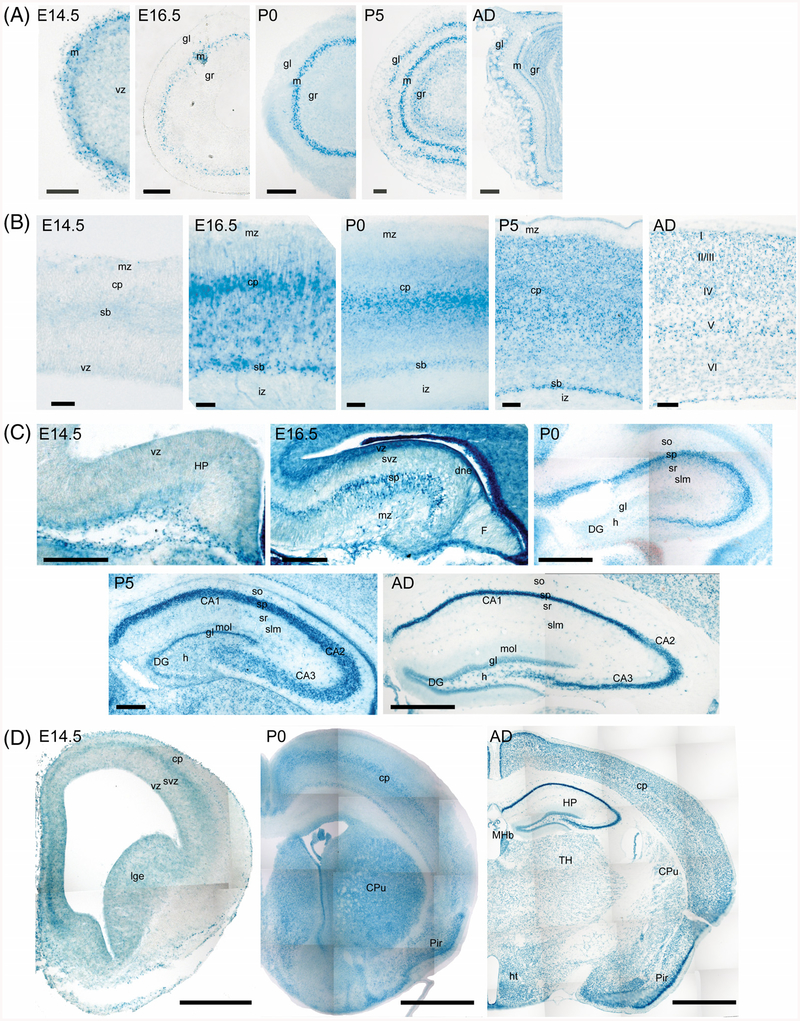
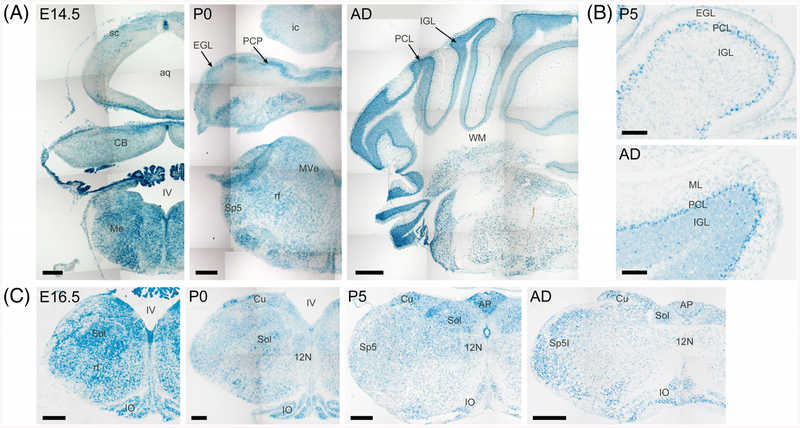
Results: Using a gene-trap mouse model for Cul5 and a knock-in-knockout mouse model for Rbx2, we show that lack of Cul5, but not Rbx2, disrupts blastocyst formation. However, Rbx2 is required for embryo survival at later embryonic stages. We also show that cul5 is expressed in the embryo proper as early as E7.5 and its expression is mostly restricted to the central nervous system and limbs at later time points. Finally, we show that rbx2 and cul5 are co-expressed in most areas of the brain during development and in the adult.

Conclusions: Our results show that Cul5, but not Rbx2, is required during early embryogenesis and suggests that Cul5 has Rbx2-independent functions in early development. In the brain, Cul5 and Rbx2 are expressed in a similar fashion, allowing the nucleation of an active CRL5 complex. Developmental Dynamics 247:1227-1236, 2018. © 2018 Wiley Periodicals, Inc.

Keywords: cul5; rbx2; CRL5; Embryogenesis; brain development.


Figures

Fig. 1.
Fig. 1.
Disruption of cul5 and rbx2 causes embryonic lethality at different developmental stages. Diagram of cul5GT (A) and rbx2GT (B) alleles indicating the LacZ cassette insertion in the first intron in both genes. Notice that whereas cul5GT and rbx2GT generate null alleles, rbx2GT can be converted to a conditional allele (rbx2fl) via FLPe recombination and to a knockout allele (rbx2KO) via CRE recombination. White boxes in exons indicate untranslated regions and gray boxes indicate coding sequences. Genotyping primers and their relative positions are indicated. Detailed explanation of genotyping strategy can be found in the Material and Methods section. C: No homozygous cul5GT embryos were collected from n = 6 litters at E3.5 and n = 5 litters analyzed at P0. On the contrary, homozygous rbx2KO embryos were collected at E3.5 (n = 15 litters) but failed to survive until birth (n = 6 litters) (D). The number of embryos obtained per genotype and age is indicated in each case. Differences from expected Mendelian ratio were tested using the Chi-square test (χ2), and P values are indicated in each case. ß-gal, beta-galactosidase; ß-geo, beta-galactosidase + Neomycin resistance gene; Chr9, chromosome 9; En2 intr1, partial Engrailed 2 Intron 1; En2 SA, Engrail 2 splicing acceptor; NeoR, Neomycin-resistance gene; pA, polyadenylation site; SA, splicing acceptor; T2A, thosea asigna virus 2A peptide.
Fig. 2.
Fig. 2.
Expression of cul5 in whole mouse embryo during mid-gestation. In toto LacZ staining at E7.5 (A), E9.5 (B, frontal view; C, lateral view), and E11.5 (D, frontal view; E, lateral view). cul5 was ubiquitously expressed in the embryo proper at E7.5 (A). By E9.5, its expression was increasingly restricted to the CNS and developing limbs (B-E). as, aortic sac; er, extraembryonic body; e, eye; ep, embryo proper; fb, left forelimb bud; hb, left hindlimb bud; III, third ventricle; ma, mandibular compartment of first branchial arch; mb, midbrain; ne, neuroepithelium; nt, neural tube; oc, otocyst; rb, rhombic lip; tv, telencephalic vesicle; te, telencephalon. Scale bar A = 50 μm. Scale bars B,C = 500 μm. Scale bars D,E = 1 mm.
Fig. 3.
Fig. 3.
Expression of cul5 in the forebrain during development and in the adult. Ontogenic analysis of cul5 expression using x-Gal staining in the olfactory bulb (A), neocortex (B), hippocampus (C), and forebrain (D) of heterozygous cul5GT animals. cul5 was strongly expressed in several areas of the forebrain, including the mitral layer in the olfactory bulb (A) and the neocortex (B). Whereas pyramidal cells of the hippocampus were positive for LacZ during development and in the adult, granule cells in the dentate gyrus expressed cul5 only in postnatal stages. CA1-CA3, Cornu Ammonis 1–3; cp, cortical plate; CPu, caudate-putamen; DG, dentate gyrus; dne, dentate neuroepithelium; F, fimbria; gl, glomerular layer; gr, granular layer; h, hilus; HP, hippocampus; ht, hypothalamus; I-VI, layer I-VI of the neocortex; iz, intermediate zone; lge, lateral ganglionic eminence; m, mitral layer; MHb, medial habenula; mol, molecular layer of the hippocampus; mz, marginal zone; Pir, piriform cortex; sb, subplate; slm, stratum lacunosum moleculare; so, stratum oriens; sp, stratum pyramidale; sr, stratum radiatum; svz, subventricular zone; vz, ventricular zone. Scale bars A,B = 50 μm. Scale bar C = 200 μm. Scale bar E = 500 μm.
Fig. 4.
Fig. 4.
Expression of cul5 in the diencephalon and medulla. cul5 was strongly expressed in the medulla at early embryonic stages (A, E14.5; C, E16.5) and its expression decreased at later time points (A,C). Expression of cul5 was strong in Purkinje cells during development and in the adult (A). High-power images (B) showed that cul5 was also expressed in the granule cells of the cerebellum, particularly in the adult (AD). AP, area postrema; aq, aqueduct; CB, cerebellum; Cu, cuneate nucleus; EGL, external granular layer; ic, inferior colliculus; IGL, internal granular layer; IO, inferior olive; IV, fourth ventricle; Me, medulla; ML, molecular layer; MVe, medial vestibular nucleus; PCP, Purkinje cell plate; PCL, Purkinje cell layer; rf, reticular formation; sc, superior colliculus; Sol, solitary tract nucleus; Sp5, spinal trigeminal nucleus; WM, white matter; 12N, hypoglossal nerve. Scale bars = 500 μm.
Fig. 5.
Fig. 5.
Expression of rbx2 during development and in the adult brain. rbx2 expression parallels cux5 expression in all the areas analyzed. In the olfactory bulb, rbx2 was strongly expressed in the mitral layer at all ages analyzed, and expression of rbx2 was detected in granule cells at postnatal stages only (A). rbx2 expression in the neocortex was detected in proliferative zones as well as in somatic areas (B) (E16.5 and P0). In the adult neocortex, strong LacZ staining was observed in all cortical layers (AD). Similar to cu/5, rbx2 was detected in proliferative areas of the hippocampus and in the stratum pyramidale at early stages (C) (E16.5). LacZ signal was detected in the dentate gyrus starting at postnatal stages and reaching maximum expression in the adult (P0 and AD). Comparative analysis of rbx2 expression in the forebrain indicated that rbx2 was ubiquitously expressed during development and in the adult (D). In the hindbrain, rbx2 expression was detected principally in Purkinje cells, deep cerebellar nuclei, and medulla (E14.5 and P0). In the adult cerebellum, a strong LacZ staining was observed in Purkinje cells, but especially in granule cells of the internal granular layer, similar to cu/5. th, thalamus; 6N, abducens nucleus; 7N, facial nucleus. Scale bars A,C = 100 μm. Scale bars D,E = 500 μm.
Fig. 6.
Fig. 6.
Co-expression of Cul5 and Rbx2 in the adult brain. Comparative analysis of Cul5 and Rbx2 by immunofluorescence in consecutive wild-type adult brain sections. In the cerebellum, both Cul5 and Rbx2 were detected in the nuclei of Purkinje cells (purple arrowheads) and broadly distributed in the internal granular layer (A). In the hippocampus, Cul5 was detected principally in the cytoplasm of the pyramidal cells of the cornu ammonis and in mossy fibers. Rbx2 was detected in the nuclei of pyramidal cells and in mossy fibers (B). In the neocortical neurons, Cul5 was detected in the cytoplasm and Rbx2 in the nucleus. On the contrary, in the caudate-putamen, both Cul5 and Rbx2 have a nuclear localization. D-D: Immunofluorescence against ß-galactosidase and Cul5 in adult rbx2GT tissue shows ubiquitous colocalization, including the neocortex (D), caudate-putamen (D”), hippocampus (D“), and cerebellum (D”’). MF, mossy fibers; sl, stratum lucidum. Scale bars A,B = 100 μm. Scale bar C = 500 μm. Scale bar D = 25 μm.

References

    1. Arai T, Kasper JS, Skaar JR, Ali SH, Takahashi C, DeCaprio JA. 2003. Targeted disruption of p185/Cul7 gene results in abnormal vascular morphogenesis. Proc Natl Acad Sci U S A 100: 9855–9860. - PMC - PubMed
    1. Bayer SA. 1983. 3H-thymidine-radiographic studies of neurogenesis in the rat olfactory bulb. Exp Brain Res 50:329–340. - PubMed
    1. Ceremuga TE, Yao XL, McCabe JT. 2001. Vasopressin-activated calcium-mobilizing (VACM-1) receptor mRNA is present in peripheral organs and the central nervous system of the laboratory rat. Endocr Res 27:433–445. - PubMed
    1. Ceremuga TE, Yao XL, McCabe JT. 2003. Cullin-5 is ubiquitous in the rat brain. Neurosci Lett 345:121–125. - PubMed
    1. Dealy MJ, Nguyen KV, Lo J, Gstaiger M, Krek W, Elson D, Arbeit J, Kipreos ET, Johnson RS. 1999. Loss of Cul1 results in early embryonic lethality and dysregulation of cyclin E. Nat Genet 23:245–248. - PubMed

Publication types

LinkOut - more resources