

Relationships Between Human-Extracted MRI Tumor Phenotypes of Breast Cancer and Clinical Prognostic Indicators Including Receptor Status and Molecular Subtype
- PMID: 30270031
- PMCID: PMC6387644
- DOI: 10.1067/j.cpradiol.2018.08.003
Relationships Between Human-Extracted MRI Tumor Phenotypes of Breast Cancer and Clinical Prognostic Indicators Including Receptor Status and Molecular Subtype
Abstract
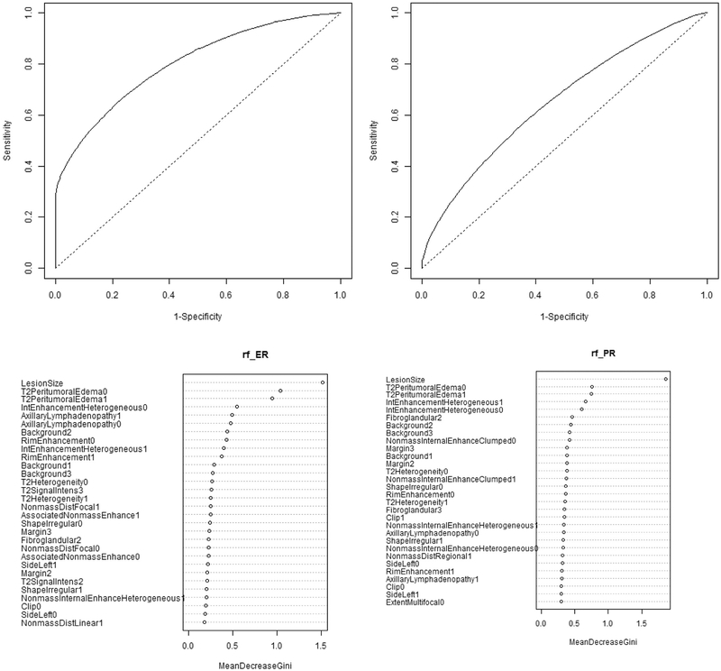
Purpose: The purpose of this study was to investigate if human-extracted MRI tumor phenotypes of breast cancer could predict receptor status and tumor molecular subtype using MRIs from The Cancer Genome Atlas project.
Materials and methods: Our retrospective interpretation study utilized the analysis of HIPAA-compliant breast MRI data from The Cancer Imaging Archive. One hundred and seven preoperative breast MRIs of biopsy proven invasive breast cancers were analyzed by 3 fellowship-trained breast-imaging radiologists. Each study was scored according to the Breast Imaging Reporting and Data System lexicon for mass and nonmass features. The Spearman rank correlation was used for association analysis of continuous variables; the Kruskal-Wallis test was used for associating continuous outcomes with categorical variables. The Fisher-exact test was used to assess correlations between categorical image-derived features and receptor status. Prediction of estrogen receptor (ER), progesterone receptor, human epidermal growth factor receptor, and molecular subtype were performed using random forest classifiers.
Results: ER+ tumors were associated with the absence of rim enhancement (P = 0.019, odds ratio [OR] 5.5), heterogeneous internal enhancement (P = 0.02, OR 6.5), peritumoral edema (P = 0.0001, OR 10.0), and axillary adenopathy (P = 0.04, OR 4.4). ER+ tumors were smaller than ER- tumors (23.7 mm vs 29.2 mm, P = 0.02, OR 8.2). All of these variables except the lack of axillary adenopathy were also associated with progesterone receptor+ status. Luminal A tumors (n = 57) were smaller compared to nonLuminal A (21.8 mm vs 27.5 mm, P = 0.035, OR 7.3) and lacked peritumoral edema (P = 0.001, OR 6.8). Basal like tumors were associated with heterogeneous internal enhancement (P = 0.05, OR 10.1), rim enhancement (P = 0.05, OR6.9), and perituomral edema (P = 0.0001, OR 13.8).
Conclusions: Human extracted MRI tumor phenotypes may be able to differentiate those tumors with a more favorable clinical prognosis from their more aggressive counterparts.
Copyright © 2018 Elsevier Inc. All rights reserved.
Figures
References
-
- Bagaria SP, et al. Personalizing breast cancer staging by the inclusion of ER, PR, and HER2. JAMA Surg 2014;149 125–9. - PubMed
-
- Huber KE, Carey LA, Wazer DE. Breast cancer molecular subtypes in patients with locally advanced disease: impact on prognosis, patterns of recurrence, and response to therapy Seminars in Radiation Oncology. Elsevier; 2009. - PubMed
-
- Wiechmann L, et al. Presenting features of breast cancer differ by molecular sub-type. Ann Surg Oncol 2009;16:2705–10. - PubMed
-
- Nguyen PL, et al. Breast cancer subtype approximated by estrogen receptor, progesterone receptor, and HER-2 is associated with local and distant recurrence after breast-conserving therapy. J Clin Oncol 2008;26:2373–8. - PubMed
MeSH terms
Substances
Grants and funding
LinkOut - more resources
Full Text Sources
Medical
Research Materials
