. 2018 Oct 18;175(3):809-821.e19.
doi: 10.1016/j.cell.2018.08.046. Epub 2018 Sep 27.

Protein AMPylation by an Evolutionarily Conserved Pseudokinase

Affiliations

Protein AMPylation by an Evolutionarily Conserved Pseudokinase

Anju Sreelatha et al. Cell. .

Abstract

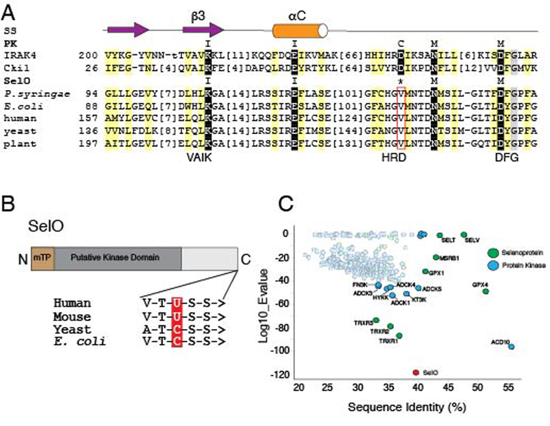
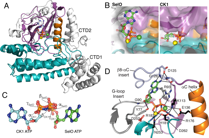
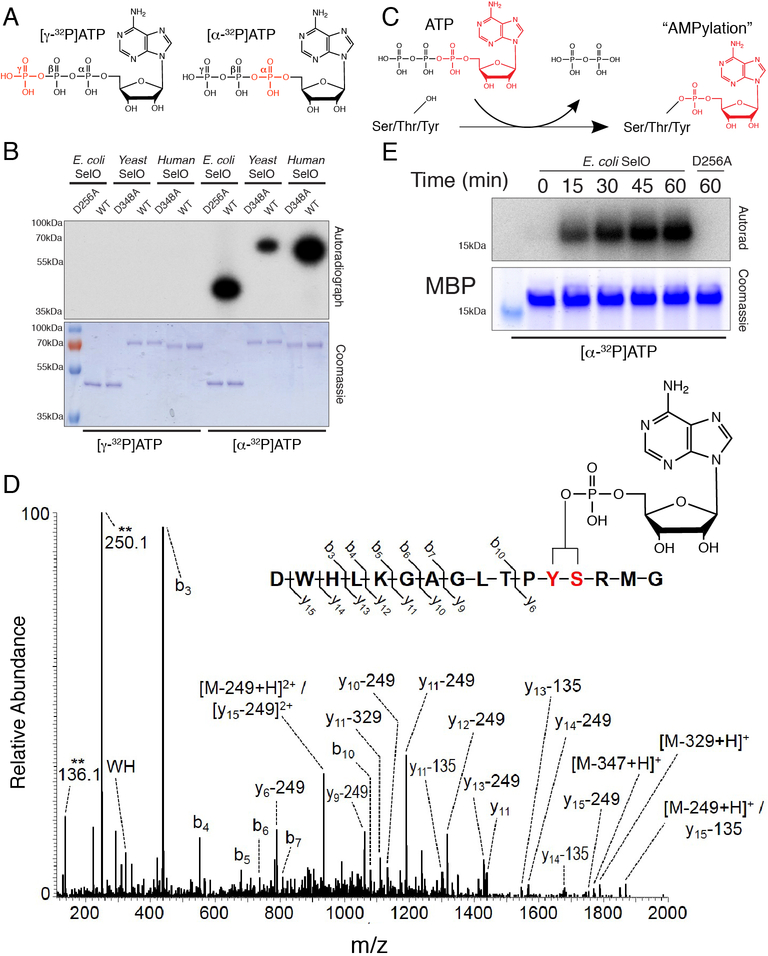
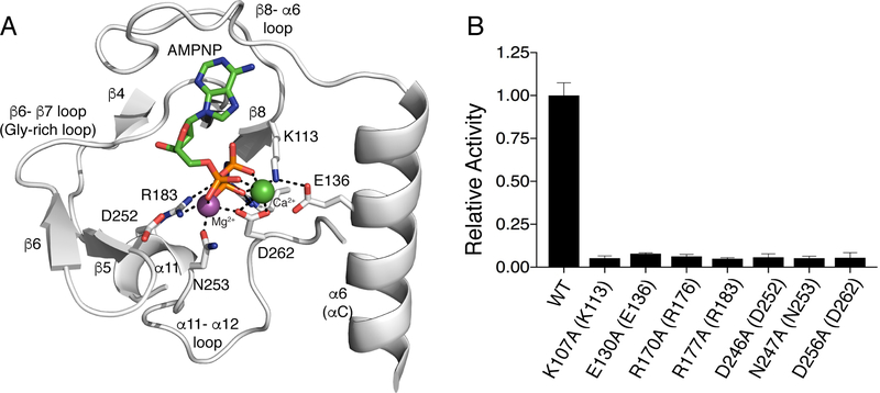
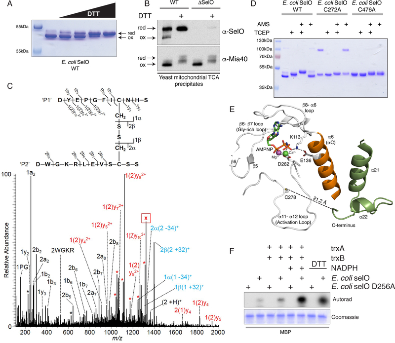
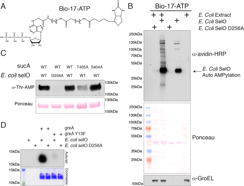
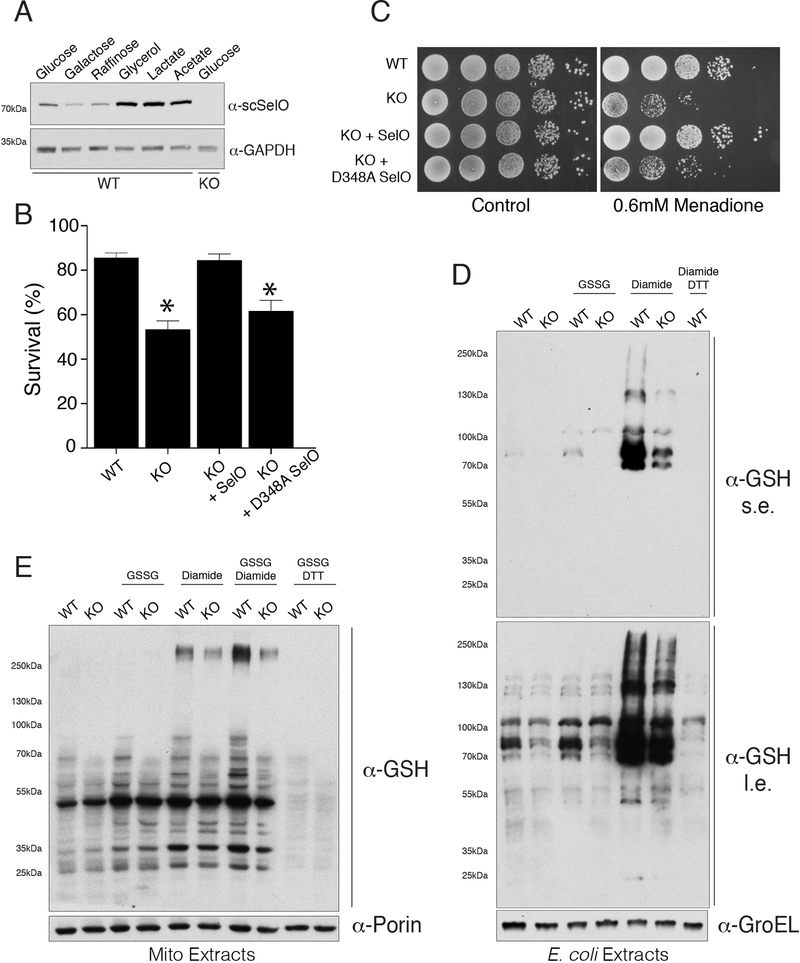
Approximately 10% of human protein kinases are believed to be inactive and named pseudokinases because they lack residues required for catalysis. Here, we show that the highly conserved pseudokinase selenoprotein-O (SelO) transfers AMP from ATP to Ser, Thr, and Tyr residues on protein substrates (AMPylation), uncovering a previously unrecognized activity for a member of the protein kinase superfamily. The crystal structure of a SelO homolog reveals a protein kinase-like fold with ATP flipped in the active site, thus providing a structural basis for catalysis. SelO pseudokinases localize to the mitochondria and AMPylate proteins involved in redox homeostasis. Consequently, SelO activity is necessary for the proper cellular response to oxidative stress. Our results suggest that AMPylation may be a more widespread post-translational modification than previously appreciated and that pseudokinases should be analyzed for alternative transferase activities.

Keywords: SELENOO; adenylylation; glutaredoxin; glutathionylation; kinase structure; oxidative stress; selenocysteine.


Conflict of interest statement

Declaration of Interests

The authors declare no competing interests.

Figures

Figure 1.
Figure 1.. SelO is an evolutionarily conserved pseudokinase. (See also Figure S1)
(A) Multiple sequence alignment highlighting conserved active site residues in the SelO pseudokinases. Conserved positions in SelO and protein kinases (PK) are highlighted yellow (hydrophobic) and gray (small). Conserved catalytic motif residues are highlighted black and labeled above: ion pair (I; VAIK), catalytic (C, HRD) and Mg2+-binding (M, DFG). Starting residue numbers are indicated before the alignment, with omitted residue numbers in brackets. Secondary structure (SS) elements are indicated above the alignment as arrow (strand) and cylinder (helix). Cki1, S. pombe protein kinase CK1; IRAK4, human interleukin-1 receptor-associated kinase 4. (B) Schematic representation of the SelO protein depicting the predicted mitochondrial targeting peptide (mTP) and kinase domain. The amino acid sequences at the C-terminus of the human, mouse, yeast and E. coli proteins are shown, highlighting the Sec (U) in the human and mouse proteins and Cys (C) in the yeast and E. coli proteins. > denotes the C-terminus. (C) BLAST analysis depicting the closest bacterial homologs retrieved from a search using the human protein kinases (blue) and selenoproteins (green) as queries. SelO is in red.
Figure 2.
Figure 2.. The crystal structure of P. syringae SelO reveals an atypical protein kinase fold with ATP flipped in the active site. (See also Figure S2 & Table S1)
(A) Ribbon representation of P. syringae SelO. The N- and C-lobes are shown in magenta and teal, respectively. The αC helix is in orange. The N-terminal extension and C-terminal domains are in white. The AMP-PNP is shown in stick representation and the Mg2+ and Ca2+ ions are shown as yellow and green spheres, respectively. (B) Surface representations illustrating the orientation of the nucleotide in the active site of P. syringae SelO (left), colored as above, but with N-lobe insertions shown in white, and protein kinase CK1 (right, pdb: 1csn) in the same orientation as SelO. (C) Ball-and-stick representation of SelO and CK1 nucleotides superimposed as a result of superposition of the proteins. The α, β, and γ phosphates of SelO and CK1 are highlighted. (D) Enlarged image of the nucleotide-binding pocket of P. syringae SelO highlighting the flipped ATP binding pocket. Two unique SelO insertions bind the flipped nucleotide, including a G90 and Y77 from the elongated G-loop (gray) that form hydrogen bonds (black dotted lines) with the ribose ring and R93 that forms hydrogen bonds with the β-phosphate, as well as D125 and G126 from the β8-αC insert (light blue) forming hydrogen bonds with the nucleotide. Two Arg sidechains (R176 and R183) form a unique charged pocket for the γ-phosphate, with the sidechains from R183 and H181 replacing the canonical ATP nucleotide binding site.
Figure 3.
Figure 3.. SelO pseudokinases AMPylate protein substrates. (See also Figure S3)
(A) Structure of the ATP molecule highlighting the position of the 32P on the γ-phosphate (left) or α-phosphate (right) in red. (B) Autoradiograph depicting the incorporation of γ32P from [γ32P]ATP (left) or α−32P from [α−32P]ATP (right) using E. coli, S. cerevisiae and human SelO (U667C), or catalytically inactive mutants. Reaction products were resolved by SDS-PAGE and visualized by Coomassie blue staining (lower) and autoradiography (upper). (C) Proposed reaction catalyzed by the SelO pseudokinases. (D) MS/MS spectrum of an AMPylated E. coli SelO peptide ion. The precursor ion, m/z 1059.47 (2+), of the AMPylated peptide was subjected to HCD fragmentation to generate the spectrum shown. Fragment ions containing the modified residue show characteristic mass shifts corresponding to loss of the AMP group (−329, −249, and −135 Da). Unique ions corresponding to neutral loss of the AMP group (labeled with **) are present at 136.1 and 250.1 Da. Location of the AMP group on the peptide can be localized to either the tyrosine or serine residue highlighted in red. (E) Time dependent incorporation of α−32P from [α−32P]ATP into MBP by SelO or SelO D256A. Reaction products were analyzed as in (B).
Figure 4.
Figure 4.. A unique active site architecture in P. syringae SelO facilitates ATP binding and AMPylation activity. (See also Figure S7).
(A) Enlarged image of the nucleotide-binding pocket of P. syringae SelO showing the detailed molecular interactions important for nucleotide binding and catalysis. Interactions are shown as dashed lines. The AMP-PNP molecule is shown in stick and the Mg2+ and Ca2+ ions are shown as purple and green spheres, respectively. (B) Activity of E. coli SelO or active site mutants using MBP and [α−32P]ATP as substrates. Reaction products were resolved by SDS-PAGE and radioactive gel bands were excised and quantified by scintillation counting. The numbering in parentheses corresponds to the residues in P. syringae SelO.
Figure 5.
Figure 5.. SelO activity is regulated by an intramolecular disulfide bridge. (See also Figure S4).
(A) Non-reducing SDS PAGE and Coomassie blue staining analysis of recombinant E. coli SelO purified in the absence of reducing agent and incubated with increasing concentrations of DTT (0 – 1mM). (B) Non-reducing SDS PAGE and protein immunoblotting of yeast mitochondrial TCA precipitates depicting endogenous S. cerevisiae SelO and Mia40. TCA precipitates were treated with or without DTT prior to electrophoresis. (C) MS/MS spectrum of E. coli SelO peptides linked by a disulfide bond: DYEPGFICNHS / DWGKRLEVSCSS. The precursor ion, m/z 1323.07 (2+) (labeled with “X”), was subjected to HCD fragmentation to generate the spectrum shown. Peaks labeled with an asterisk (*) correspond to neutral loss of ammonia (−17 Da) or water (−18 Da) from fragment ions. The peak labels are color coded depending on the status of the disulfide bond (SS) for that particular ion: black (no SS), red (intact SS), and blue (asymmetric cleavage of SS, α or β). (D) Non-reducing SDS PAGE and Coomassie blue staining analysis of recombinant E. coli SelO or the C272A and C476A mutants purified under non-reducing conditions and incubated with the reducing agent TCEP or the alkylating agent AMS. The species at ~100kDa in the SelO C272A mutant is a dimer formed between two molecules of E. coli SelO linked by an intermolecular disulfide. (E) Enlarged image of the active site highlighting the activation loop C278 (C272 in E. coli SelO) and the C-terminus of the protein. The α6 (αC equivalent) is in orange and the α21 and α22 helices are in green. The AMP-PNP molecule is shown in stick and the Mg2+ and Ca2+ ions are shown as purple and green spheres, respectively. (F) Incorporation of 32P AMP from [α−32P]ATP by E. coli SelO or the D256A mutant under non-reducing or reducing conditions (DTT or the thioredoxin system). (trxA; E. coli thioredoxin, trxB; E. coli thioredoxin reductase). Reaction products were analyzed as in Figure 3B.
Figure 6.
Figure 6.. SelO AMPylates mitochondrial proteins. (See also Figure S5 & Table S2).
(A) Structure of biotin-17-ATP (bio-17-ATP) used in these experiments to identify SelO substrates. (B) Representative blot using avidin-HRP to detect biotinylated proteins following incubation of E. coli extracts with bio-17-ATP and E. coli SelO or the D256A mutant. The Ponceau stained membrane and an immunoblot for GroEL are shown as loading controls. (C) α-Thr AMP protein immunoblotting of Ni-NTA affinity purified His-tagged sucA (or mutants) from SelO KO E.coli extracts expressing untagged WT SelO or the inactive mutant. The Ponceau stained membrane is shown. (D) Autoradiograph depicting the incorporation of α−32P AMP from [α−32P]ATP by E. coli SelO or the D256A mutant into E. coli grxA. Reaction products were analyzed as in Figure 3B.
Figure 7.
Figure 7.. SelO protects yeast cells from oxidative stress and regulates protein S-glutathionylation. (See also Figure S6).
(A) Representative protein immunoblots of WT and SelO KO yeast cell extracts (strain MR6) grown in medium with the indicated carbon source. S. cerevisiae SelO (ScSelO) and GAPDH (loading control) are shown. (B) Percent survival of S. cerevisiae (strain MR6) WT, SelO KO or SelO KO cells complemented with WT or D348A SelO following treatment with 100 μM H2O2 in glucose minimal medium for 200 min at 28°C. Results represent the mean of 3 independent experiments. * p < 0.005 vs WT. (C) Representative growth assays of S. cerevisiae (strain BY4741) untreated (left) or 0.6 mM menadione treated (right). WT, SelO KO or SelO KO complemented with WT or D348A SelO strains were analyzed. (D) Representative protein immunoblots of WT and SelO KO E. coli extracts following treatment of intact cells with oxidized glutathione (GSSG) or diamide. Cells were also treated with diamide followed by DTT as a negative control. Extracts were probed with anti-glutathionylation (GSH) and E. coli GroEL (loading control). Results are representative of at least 3 independent experiments. s.e. (short exposure). l.e. (long exposure). (E) Representative protein immunoblots of crude mitochondrial extracts isolated from WT and SelO KO S. cerevisiae (strain MR6) following treatment of intact mitochondria with oxidized glutathione (GSSG) and/or diamide. Mitochondria were also treated with GSSG followed by DTT as a negative control. Extracts were probed with anti-glutathionylation (GSH) and S. cerevisiae porin (loading control). Results are representative of at least 3 independent experiments.

Comment in

References

    1. Adams PD, Afonine PV, Bunkoczi G, Chen VB, Davis IW, Echols N, Headd JJ, Hung LW, Kapral GJ, Grosse-Kunstleve RW, et al. (2010). PHENIX: a comprehensive Python-based system for macromolecular structure solution. Acta Crystallogr D Biol Crystallogr 66, 213–221. - PMC - PubMed
    1. Altschul SF, Gish W, Miller W, Myers EW, and Lipman DJ (1990). Basic local alignment search tool. J Mol Biol 215, 403–410. - PubMed
    1. Altschul SF, Madden TL, Schaffer AA, Zhang J, Zhang Z, Miller W, and Lipman DJ (1997). Gapped BLAST and PSI-BLAST: a new generation of protein database search programs. Nucleic Acids Res 25, 3389–3402. - PMC - PubMed
    1. Bailey FP, Byrne DP, McSkimming D, Kannan N, and Eyers PA (2015). 2) Going for broke: targeting the human cancer pseudokinome. Biochem J 466, 201. - PubMed
    1. Borek D, Cymborowski M, Machius M, Minor W, and Otwinowski Z (2010). Diffraction data analysis in the presence of radiation damage. Acta Crystallogr D Biol Crystallogr 66, 426–436. - PMC - PubMed

Publication types

MeSH terms