. 2018 Oct 18;72(2):355-368.e4.
doi: 10.1016/j.molcel.2018.08.021. Epub 2018 Sep 27.

RIG-I Uses an ATPase-Powered Translocation-Throttling Mechanism for Kinetic Proofreading of RNAs and Oligomerization

Affiliations

RIG-I Uses an ATPase-Powered Translocation-Throttling Mechanism for Kinetic Proofreading of RNAs and Oligomerization

Swapnil C Devarkar et al. Mol Cell. .

Abstract

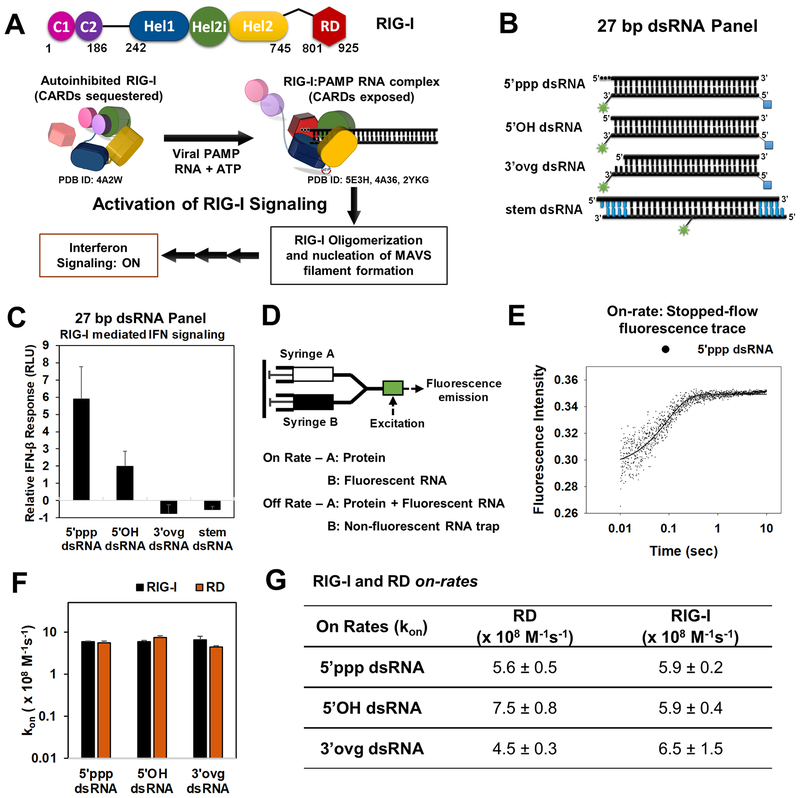
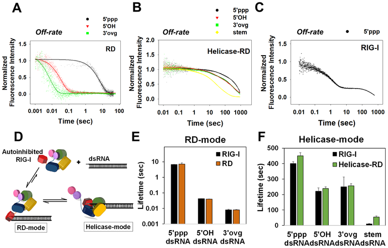
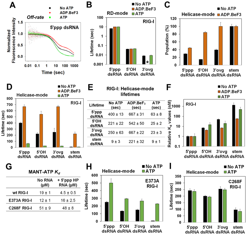
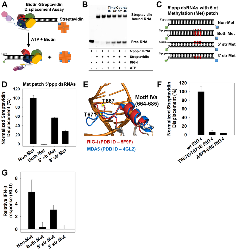
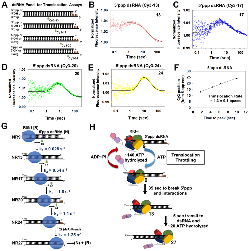
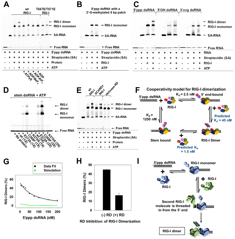
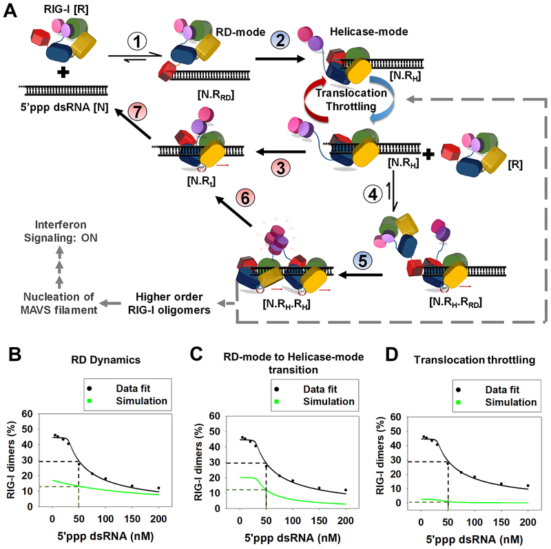
RIG-I has a remarkable ability to specifically select viral 5'ppp dsRNAs for activation from a pool of cytosolic self-RNAs. The ATPase activity of RIG-I plays a role in RNA discrimination and activation, but the underlying mechanism was unclear. Using transient-state kinetics, we elucidated the ATPase-driven "kinetic proofreading" mechanism of RIG-I activation and RNA discrimination, akin to DNA polymerases, ribosomes, and T cell receptors. Even in the autoinhibited state of RIG-I, the C-terminal domain kinetically discriminates against self-RNAs by fast off rates. ATP binding facilitates dsRNA engagement but, interestingly, makes RIG-I promiscuous, explaining the constitutive signaling by Singleton-Merten syndrome-linked mutants that bind ATP without hydrolysis. ATP hydrolysis dissociates self-RNAs faster than 5'ppp dsRNA but, more importantly, drives RIG-I oligomerization through translocation, which we show to be regulated by helicase motif IVa. RIG-I translocates directionally from the dsRNA end into the stem region, and the 5'ppp end "throttles" translocation to provide a mechanism for threading and building a signaling-active oligomeric complex.

Keywords: ATP; RIG-I; RNA discrimination; innate immunity; kinetic proofreading; kinetics; oligomerization; self-versus-non-self; translocation.


Conflict of interest statement

Declaration of Interests

The authors declare no competing interests.

Figures

Figure 1.
Figure 1.. RIG-I activation pathway and RIG-I/RD on-rates on 5’ppp and non-PAMP dsRNAs.
(A) Schematic of a general activation mechanism of RIG-I. (B) Cartoon of the dsRNAs used in the study (green asterisk-DY547/Cy3; blue square-biotin; blue bars-DNA bases). (C) IFN-β reporter activation in HEK293T cells expressing RIG-I and transfected with the dsRNA panel from B. Error bars are SEM from quadruplicate sets. (D) Stopped-flow experimental set up for on-rate and off-rate measurements. (E) Representative stopped-flow time course shows an increase in fluorescence intensity upon binding of RIG-I (45 nM) to 5’ppp dsRNA (10 nM) at 25°C. (F, G) On-rates of RIG-I and RD. The associated errors from the linear fits are shown. See also Figure S1.
Figure 2.
Figure 2.. Off-rates of 5’ppp and non-PAMP RNAs from RD, Helicase-RD, and RIG-I.
(A-C) Stopped-flow kinetics of RNA dissociation. A preformed complex of RD (A), Helicase-RD (B), or RIG-I (C) with the specified fluorophore-tagged RNA was chased with a 10-fold excess of unlabeled 5’ppp ds12 hairpin RNA at 25°C. (D) RIG-I forms two distinct complexes with dsRNA: RD-mode and Helicase-mode conformations. (E-F) Lifetimes of RD-mode (E) and Helicase-mode (F) RIG-I complexes calculated from reciprocal of corresponding off-rates. Representative kinetic traces are an average of 4–6 traces. See also Figure S2 and Table S1.
Figure 3.
Figure 3.. Off-rates of 5’ppp and non-PAMP RNAs in the presence of ADP.BeF3 and ATP.
(A) Representative off-rate kinetics of RIG-I from 5’ppp dsRNA. (B) Lifetimes of the RD-mode population. (*) indicates that an RD-mode population was not observed. Relative percentages (C) and lifetimes (D) of the Helicase-mode population of RIG-I. (E) Helicase-mode lifetimes of RIG-I:RNA complexes in the presence of the specified ATP analog (F) Kd values of RIG-I:RNA complexes normalized to the 5’ppp dsRNA:RIG-I complex. (G) MANT-ATP Kd values. (H-I) Lifetimes of the Helicase-mode population of E373A (H) and C268F RIG-I (I). Representative kinetic traces are an average of 4–6 traces. See also Figure S3 and Tables S2–S6.
Figure 4.
Figure 4.. Directionality of RIG-I translocation and role of Helicase motif IVa.
(A) Biotin-streptavidin displacement assay schematic. (B) Time-course of biotin-streptavidin displacement from 5’ppp dsRNA (25 nM) by RIG-I (25 nM). (C) 27bp methylated patch 5’ppp dsRNAs to assess directionality of RIG-I translocation (Red bars- 5nt patch of 2’-O-methylated nucleotides placed 15bp downstream of the 5’ppp end). (D) Streptavidin displacement activity on methylated patch 5’ppp dsRNAs. Error bars are SEM from triplicates. (E) Helicase motif IVa of RIG-I (red) and MDA5 (blue). T667E and T671E in RIG-I are shown as yellow circles. (F) Streptavidin displacement by motif IVa mutants on the 5’ppp dsRNA. Error bars are SEM from triplicates. (G) IFN-β reporter activity of RIGI transfected HEK293T cells upon activation by the 2’-O-methylated 5’ppp dsRNAs. Error bars are SEM from quadruplicates. See also Figure S4.
Figure 5.
Figure 5.. Stepping rates of RIG-I translocation.
(A) Schematic of a panel of Cy3 labeled 27bp dsRNAs for RIG-I translocation studies (Brown bars-DNA bases). (B-E) Transient state kinetics of RIG-I translocation on the 5’ppp dsRNA measured by Cy3 fluorescence intensity changes. The position of Cy3 from the RNA-end is denoted. Solid lines are fit to the model in G. (F) Time-to-peak increases linearly with Cy3 position and slope provides the average translocation rate. (G) The minimal translocation model fits the stopped-flow translocation data in B-E (solid lines) with the given stepping rates. (H) Translocation throttling mechanism of RIG-I at the 5’ppp end. See also Figure S5 and Table S7.
Figure 6.
Figure 6.. ATP facilitates cooperative RIG-I dimerization on 5’ppp dsRNA by 5’-end threading mechanism.
(A-B) EMSAs of RIG-I or specified mutant (25 nM) with DY547-tagged 5’ppp dsRNA (25 nM) (A) and methylation patch carrying 5’ppp dsRNA (25 nM) (B). (C) EMSAs of DY547 labeled 5’ppp dsRNA, 5’OH dsRNA and 3’ovg dsRNA (100 nM each) with RIG-I (100 nM). (D) EMSAs of Cy3 labeled stem dsRNA (100 nM) with RIG-I and specified mutants (100 and 300 nM each). (E) EMSA of DY547 labeled 5’ppp dsRNA (25 nM) with RIG-I or specified mutant (25 nM) in presence or absence of ATP. (F) Cooperativity model for RIG-I dimerization. (G) RIG-I (50 nM) was titrated with increasing concentrations of 5’ppp dsRNA (5–200 nM) with ATP (2mM) and EMSA was used to determine percent dimers. Green line represents the expected trend for non-cooperative RIG-I dimerization, and the black line shows the best fit to the cooperative dimerization model in E. (H) 5’ppp dsRNA (25 nM) and RIG-I (75 nM) were incubated with and without RD (75 nM) and ATP (2 mM), and percent RIG-I dimers from EMSA are plotted. Error bars are SEM from triplicates. (I) The 5’-end threading model is consistent with results in G. Dotted lines in EMSA gel blots represent deletion of lanes. See also Figure S6.
Figure 7.
Figure 7.. Kinetic proofreading pathway for RIG-I oligomerization and RNA discrimination.
(A) The seven steps show the kinetic pathway of RIG-I oligomerization. (1) RIG-I forms the RD-mode complex. (2) The RD-mode complex transitions into an ATPase active Helicase-mode complex that undergoes multiple rounds of ATP hydrolysis at the 5’-end (Translocation-throttling). (3) Helicase-mode complex translocates away from the dsRNA-end, or (4) binds a second molecule of RIG-I in the RD-mode. (5) Second RIG-I molecule transitions to Helicase-mode resulting in RIG-I dimer on dsRNA. (6–7) RIG-I dimer can translocate and dissociate from the RNA. Steps shaded in blue (2,5) are facilitated by ATP binding and steps shaded in red are driven by ATP hydrolysis (3,6 and 7). (B-D) The decrease in RIG-I dimers with increasing 5’ppp dsRNA concentration (Figure S6D) fit well to the kinetic proofreading pathway from A (black). The experimentally measured values used for the fitting are: Step 1 and 4 (kon: 6 × 10⁸ M−1s−1, koff: 0.15 s−1), Step 3 and 6 (0.028 s−1), step 7 (1.2 s−1); predicted rates from best fit for step 2 and 5 were 0.03 s−1 and 0.06 s−1, respectively. Simulations (green) were carried out by either substituting the RD-mode off-rates of 5’ppp dsRNA in the model with 3’ovg dsRNA off-rate (B), decreasing the N.RRD to N.RH transition rate by 3-fold (C), and removing all translocation throttling steps at the 5’ end for monomer and dimer (D).

References

    1. Bamming D, and Horvath CM (2009). Regulation of signal transduction by enzymatically inactive antiviral RNA helicase proteins MDA5, RIG-I, and LGP2. J Biol Chem 284, 9700–9712. - PMC - PubMed
    1. Banerjee K, Kolomeisky AB, and Igoshin OA (2017). Accuracy of Substrate Selection by Enzymes Is Controlled by Kinetic Discrimination. J Phys Chem Lett 8, 1552–1556. - PubMed
    1. Blanchard SC, Gonzalez RL, Kim HD, Chu S, and Puglisi JD (2004). tRNA selection and kinetic proofreading in translation. Nat Struct Mol Biol 11, 1008–1014. - PubMed
    1. Devarkar SC, Wang C, Miller MT, Ramanathan A, Jiang F, Khan AG, Patel SS, and Marcotrigiano J (2016). Structural basis for m7G recognition and 2’-O-methyl discrimination in capped RNAs by the innate immune receptor RIG-I. Proc Natl Acad Sci U S A 113, 596–601. - PMC - PubMed
    1. Dillingham MS, Wigley DB, and Webb MR (2000). Demonstration of unidirectional single-stranded DNA translocation by PcrA helicase: measurement of step size and translocation speed. Biochemistry 39, 205–212. - PubMed

Publication types

MeSH terms

Supplementary concepts

LinkOut - more resources