. 2018 Dec 4;26(12):1604-1611.e4.
doi: 10.1016/j.str.2018.08.007. Epub 2018 Sep 27.

N-Terminal Domains of Cardiac Myosin Binding Protein C Cooperatively Activate the Thin Filament

Affiliations

N-Terminal Domains of Cardiac Myosin Binding Protein C Cooperatively Activate the Thin Filament

Cristina Risi et al. Structure. .

Abstract

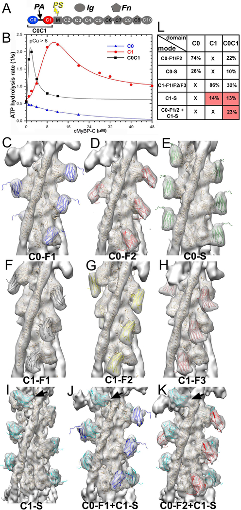
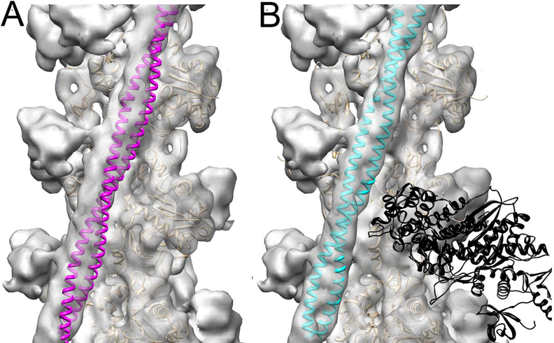
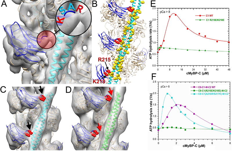
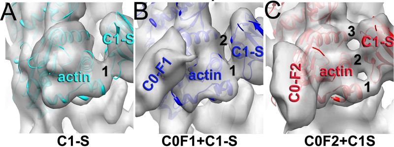
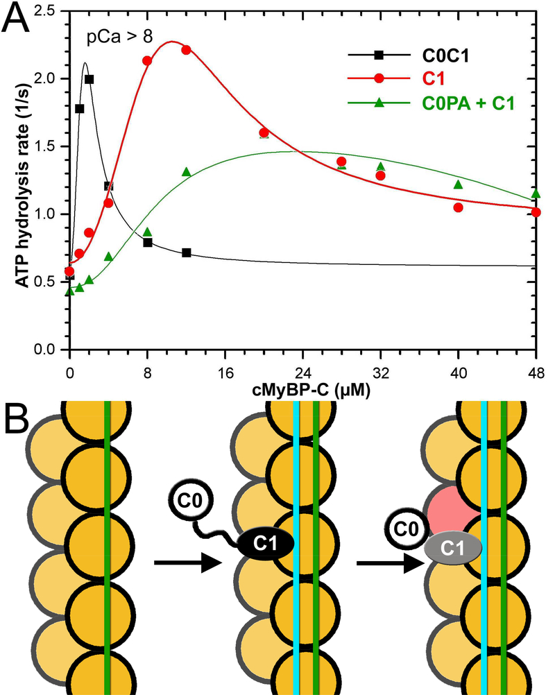
Muscle contraction relies on interaction between myosin-based thick filaments and actin-based thin filaments. Myosin binding protein C (MyBP-C) is a key regulator of actomyosin interactions. Recent studies established that the N'-terminal domains (NTDs) of MyBP-C can either activate or inhibit thin filaments, but the mechanism of their collective action is poorly understood. Cardiac MyBP-C (cMyBP-C) harbors an extra NTD, which is absent in skeletal isoforms of MyBP-C, and its role in regulation of cardiac contraction is unknown. Here we show that the first two domains of human cMyPB-C (i.e., C0 and C1) cooperate to activate the thin filament. We demonstrate that C1 interacts with tropomyosin via a positively charged loop and that this interaction, stabilized by the C0 domain, is required for thin filament activation by cMyBP-C. Our data reveal a mechanism by which cMyBP-C can modulate cardiac contraction and demonstrate a function of the C0 domain.

Keywords: cardiac muscle; cryoelectron microscopy; myosin binding protein C; thin filament.


Conflict of interest statement

Declaration of Interests: The authors declare no competing interests.

Figures

Figure 1.
Figure 1.
3D reconstructions and corresponding frequencies of structural classes revealed in the C0C1 decorated TFs. (A) cMyBP-C is composed of 8 Immunoglobulin (Ig)-like and 3 fibronectin (Fn)-like domains numbered C0 through C10 starting consecutively from the N-terminus to the C-terminus of the molecule. C0 (blue) and C1 (red) domains are connected by a flexible proline-alanine (PAL) linker, while the M-domain which links C1 and C2 contains functionally significant phosphorylation sites (PS). (B) Effects of C0 (blue curve), C1 (red curve) and tandem C0-C1 (black curve) on TF-dependent myosin-S1 ATPase activity at pCa>8. (C-K) 3D reconstructions of structural classes of C0C1-decorated TFs show all the modes of C0 (C-E) and C1 (F-I) binding to the cTF observed earlier (Harris et al., 2016) along with the two structural classes where C0 and C1 bind simultaneously to the cTF (J and K). High resolution structure of C0 (pdb: 2K1M) is shown in blue (C0-F1 mode), red (C0-F2 mode), or green (C0-S mode). The high resolution structure of the C1 (pdb: 2V6H) is shown in black (C1-F1 mode), yellow (C1-F2 mode), brown (C1-F3 mode), or cyan (C1-S mode). Actin and Tm molecules are shown as tan ribbons. Reconstructions are shown as grey transparent surfaces filtered to the resolution determined for each class (Figure S1C). Interaction of C1 with Tm in the C1-S, C0-F1+C1-S, and C0-F2+C1-S are marked with black arrows. (L) Frequencies for individual classes of cTFs decorated with individual C0 and C1 domains obtained earlier (Harris et al., 2016) are compared with those calculated for the C0C1-decorated TFs. Frequencies of classes where C1 interacts with Tm are marked in red.
Figure 2.
Figure 2.
C1 binding to the Tm cable traps it in the myosin rather than Ca2+-induced c-open structural state. (A) Atomic model of the cardiac open (c-open) structural state of the cardiac TF (Protein Data Bank ID code 5NOJ) (Risi et al., 2017) was rigidly docked into the 3D reconstruction of the C1-S structural class to show that Tm (magenta ribbons) does not fit into the portion of the electron density map that corresponds to the Tm cable. (B) The model of the myosin-Tpm-F-actin complex (von der Ecken et al., 2016) (Protein Data Bank ID code 5JLH) docked into the 3D reconstruction of the C1-S structural class without any perturbations unambiguously shows that Tm (cyan ribbons) position in the 3D reconstruction of the C1-S class corresponds to the myosin state of the TF. 3D reconstructions are shown as grey transparent surfaces. Actin molecules are shown as tan ribbons, while rigor bound myosin-S1 head is shown as black ribbons.
Figure 3.
Figure 3.
R215 and K218 amino acid residues of C1 maintain its interaction with Tm which is required for the TF activation by the cMyBP-C NTD. (A) A 9 Å 3D reconstruction of the C1-S class shows a bridge of density (red circle) between C1 (blue ribbons) and Tm (cyan ribbons). Insert shows that C1 loop involved in the interaction is comprised of two positively charged (red) and two neutral (cyan) residues. (B) Residues R215 and K218 (red spheres) of C1 loop are located in proximity to negatively charged residues of Tm (yellow spheres). Tm model is from (Behrmann et al., 2012). (C-D) In the 3D reconstruction of the wild type C1 in the C1-S mode there is a bridge of density (black arrows) between C1 (C, blue ribbons) and Tm in the myosin state (C, cyan ribbons). It is absent in the 3D reconstruction of the C1-S class of the C1(R215E/K218E)-decorated TFs (D, red arrows) where Tm is in the c-closed position (D, green ribbons). (E) Effects of C1 (red curve), and C1(R215E/K218E) (green curve) on TF-dependent myosin-S1 ATPase activity at pCa>8. (F) Effects of C0C1MC2 (magenta curve), C0C1(R215E/K218E)MC2 (green curve), and C0C1(A216R/S217K)MC2 on TF-dependent myosin-S1 ATPase activity at pCa>8.
Figure 4.
Figure 4.
Effects of C0 on the interaction of C1 with F-actin. Comparison of the contacts between actin and C1 in the reconstructions of C1-S (A), C0-F1+C1-S (B) and C0-F2+C1S (C) shows that in the absence of bound C0, C1 has one contact with actin (A), while binding of C0 in the F1 and F2 models increases the number of contacts to two (B) and three (C), respectively.
Figure 5.
Figure 5.
C0 and C1 Ig-domains of cMyBP-C work in tandem to activate the cTF. (A) Effects of C0-PA-C1 (black curve), C1 (red curve), and equimolar mixture of C0-PA and C1 on TF-dependent myosin-S1 ATPase activity at pCa>8. (B) The model of activation of the cTF by C0-PA-C1 fragment of cMyBP-C. At relaxing conditions Tm is in the inactive (c-closed and c-blocked) states (Risi et al., 2017) (green line). Upon C1 binding in the C1-S mode the Tm cable is azimuthally shifted to its myosin (cyan line) state (Figure 2B) by means of interaction with the R215 and K218 residues of C1 (Figure 3). Bound C1 enforces interaction of C0 with the front of the same actin molecule which enhances the C1 interaction with F-actin (Fig. 4A-C). Actin molecules are shown in yellow. C0 Ig-domain is shown as white circle, C1 domain bound in the C1-S mode is black oval, while when activated by the C0 domain is marked in grey.

References

    1. Spudich JA, Huxley HE, and Finch JT (1972). Regulation of skeletal muscle contraction. II. Structural studies of the interaction of the tropomyosin-troponin complex with actin. J. Mol. Biol 72, 619–632. - PubMed
    1. McKillop DFA, and Geeves MA (1993). Regulation of the interaction between actin and myosin subfragment-1: evidence for three states of the thin filament. Biophys. J 65, 693–701. - PMC - PubMed
    1. Vibert P, Craig R, and Lehman W (1997). Steric-model for activation of muscle thin filaments. J. Mol. Biol 266, 8–14. - PubMed
    1. Houmeida A, Heeley DH, Belknap B, and White HD (2010). Mechanism of regulation of native cardiac muscle thin filaments by rigor cardiac myosin-S1 and calcium. J. Biol. Chem 285, 32760–32769. - PMC - PubMed
    1. Heeley DH, Belknap B, and White HD (2006). Maximal activation of skeletal muscle thin filaments requires both rigor myosin S1 and calcium. J. Biol. Chem 281, 668–676. - PubMed

Publication types

LinkOut - more resources