. 2018 Sep 9:2018:3290372.
doi: 10.1155/2018/3290372. eCollection 2018.

MicroRNA-132, Delivered by Mesenchymal Stem Cell-Derived Exosomes, Promote Angiogenesis in Myocardial Infarction

Affiliations

MicroRNA-132, Delivered by Mesenchymal Stem Cell-Derived Exosomes, Promote Angiogenesis in Myocardial Infarction

Teng Ma et al. Stem Cells Int. .

Abstract

Background: To cure ischemic diseases, angiogenesis needs to be improved by various strategies in ischemic area. Considering that microRNA-132 (miR-132) regulates endothelial cell behavior during angiogenesis and the safe and efficacious delivery of microRNAs in vivo is rarely achieved, an ideal vehicle for miR-132 delivery could bring the promise for ischemic diseases. As a natural carrier of biological molecules, exosomes are more and more developed as an ideal vehicle for miRNA transfer. Meanwhile, mesenchymal stem cells could release large amounts of exosomes. Thus, this study aimed to investigate whether MSC-derived exosomes can be used for miR-132 delivery in the treatment of myocardial ischemia.

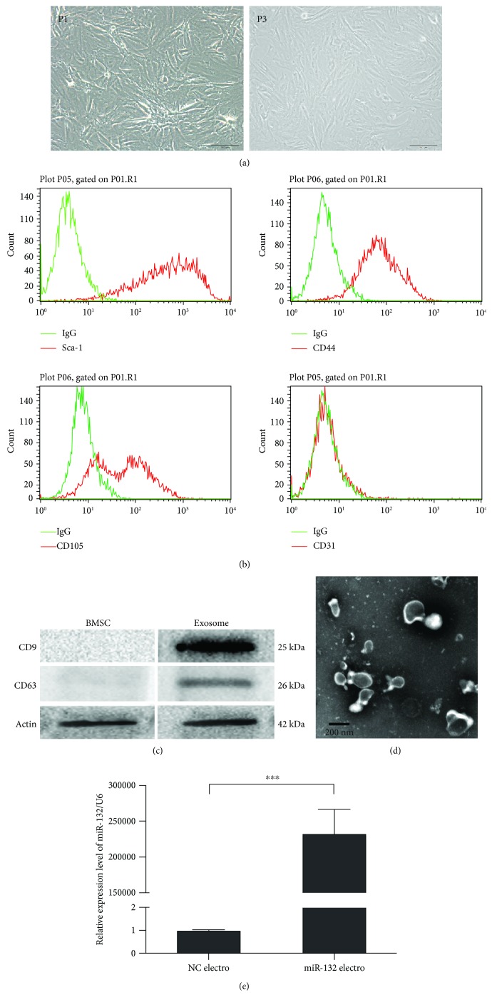
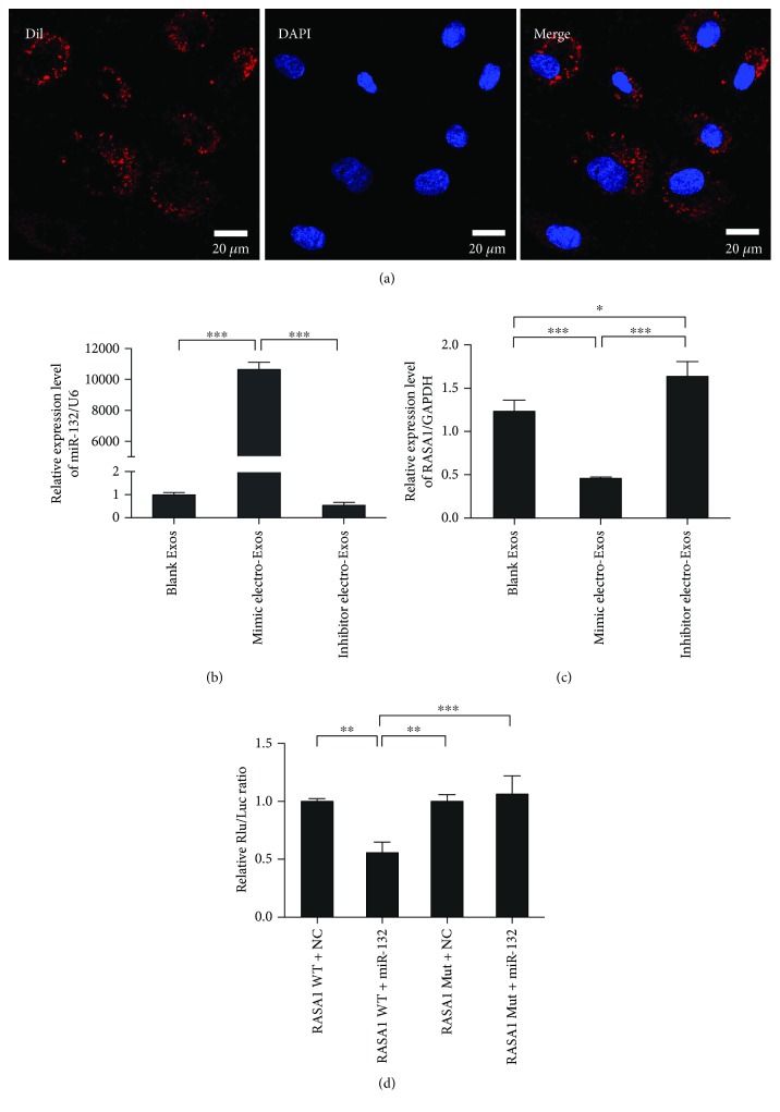
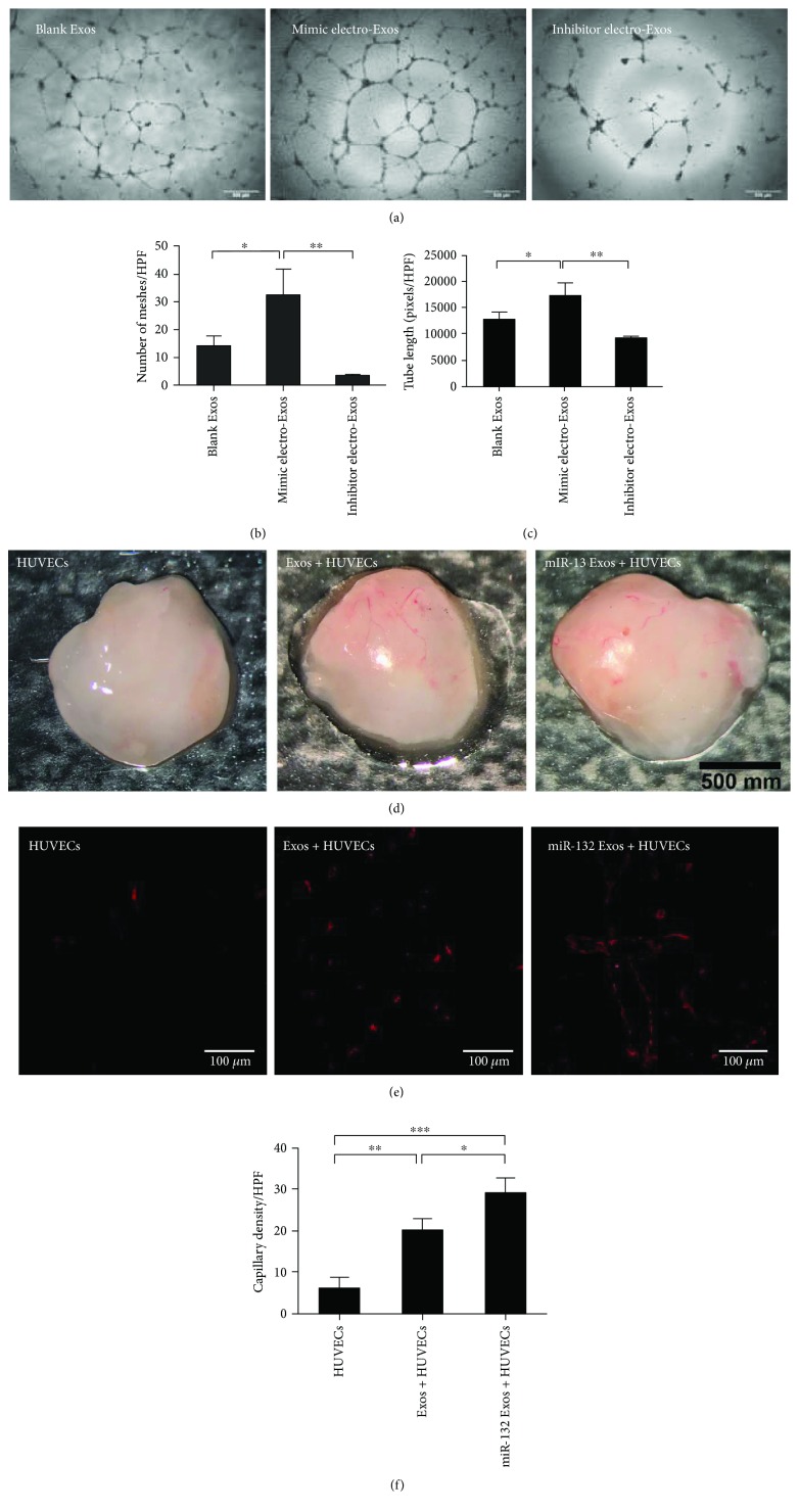
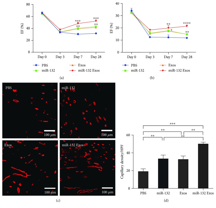
Methods: MSC-derived exosomes were electroporated with miR-132 mimics and inhibitors. After electroporation, miR-132 exosomes were labelled with DiI and added to HUVECs. Internalization of DiI-labelled exosomes was examined by fluorescent microscopy. Expression levels of miR-132 in exosomes and HUVECs were quantified by real-time PCR. The mRNA levels of miR-132 target gene RASA1 in HUVECs were quantified by real-time PCR. Luciferase reporter assay was performed to examine the targeting relationship between miR-132 and RASA1. The effects of miR-132 exosomes on the angiogenic ability of endothelial cells were evaluated by tube formation assay. Matrigel plug assay and myocardial infarction model were used to determine whether miR-132 exosomes can promote angiogenesis in vivo.

Results: miR-132 mimics were effectively electroporated and highly detected in MSC-derived exosomes. The expression level of miR-132 was high in HUVECs preincubated with miR-132 mimic-electroporated exosomes and low in HUVECs preincubated with miR-132 inhibitor-electroporated exosomes. The expression level of RASA1, miR-132 target gene, was reversely correlated with miR-132 expression in HUVECs pretreated with exosomes. Luciferase reporter assay further confirmed that RASA1 was a direct target of miR-132. Exosomes loaded with miR-132, as a vehicle for miRNA transfer, significantly increased tube formation of endothelial cells. Moreover, subcutaneous injection of HUVECs pretreated with miR-132 exosomes in nude mice significantly increased their angiogenesis capacity in vivo. In addition, transplantation of miR-132 exosomes in the ischemic hearts of mice markedly enhanced the neovascularization in the peri-infarct zone and preserved heart functions.

Conclusions: The findings suggest that the export of miR-132 via MSC-derived exosomes represents a novel strategy to enhance angiogenesis in ischemic diseases.


Figures

Figure 1
Figure 1
Characterization of BMSCs and BMSC-derived exosomes. (a) Morphology of MSCs (P1, P3) observed under an inverted fluorescence microscope. Scale bar: 100 μm. (b) Phenotypic analysis of cell surface antigens of MSCs by flow cytometry (n = 3). (c) Surface marker proteins of BMSCs and BMSC-derived exosomes analyzed by Western immunoblotting (n = 3). (d) Morphology of MSC-derived exosomes under transmission electron microscopy. Scale bar: 200 nm. (e) The expression level of miR-132 determined by Q-PCR (n = 3). ∗∗∗P < 0.001. NC: negative control.
Figure 2
Figure 2
Internalization of miR-132-electroporated exosomes and detection of target gene RASA1. (a) Confocal images of DiI-labelled exosomes taken up by HUVECs. Scale bar: 20 μm. (b, c) HUVECs were incubated with miR-132 mimics or inhibitor-electroporated exosomes for 2 h. The relative expression level of miR-132 and its target gene RASA1 was detected by RT-PCR (n = 3). (d) 293T was cotransfected with miR-132 mimics or NC and firefly luciferase reporter plasmid containing wild-type or mutant-type 3′UTR of RASA1. After incubation for 48 h, the firefly luciferase activity of each sample was detected and normalized to the Renilla luciferase activity (n = 3). The data represent the mean ± SEM of triplicates. P < 0.05, ∗∗P < 0.01, ∗∗∗P < 0.001.
Figure 3
Figure 3
miR-132-electroporated exosomes promoted angiogenesis in vitro and in vivo. (a) Tube formation assay on Matrigel was assessed 6 h after seeding HUVECs pretreated with blank, miR-132 mimic-electroporated or miR-132 inhibitor-electroporated exosomes. Scale bar: 500 μm. (b, c) Quantitative assessment of the total number of meshes and tube length (n = 3). P < 0.05, ∗∗P < 0.01. (d) Gross look of Matrigel plugs. (e, f) Immunofluorescence staining of vessels in the sections of Matrigel plugs and quantitative assessment of capillaries per high-power field in each group (n = 3). P < 0.05, ∗∗∗P < 0.001.
Figure 4
Figure 4
miR-132-electroporated exosomes preserve cardiac function and promoted angiogenesis in MI model. (a, b) Quantitative assessment of LVEF and FS value in each group after MI (n = 3). P < 0.05, ∗∗P < 0.01, ∗∗∗P < 0.001, ∗∗∗∗P < 0.0001. (c, d) Immunofluorescence staining of vessels in the sections of heart tissue and quantitative assessment of capillaries per high-power field in each group. Scale bar: 500 μm (n = 3). ∗∗P < 0.01, ∗∗∗P < 0.001.

References

    1. Katare R., Riu F., Mitchell K., et al. Transplantation of human pericyte progenitor cells improves the repair of infarcted heart through activation of an angiogenic program involving micro-RNA-132. Circulation Research. 2011;109(8):894–906. doi: 10.1161/CIRCRESAHA.111.251546. - DOI - PMC - PubMed
    1. Wen Z., Huang W., Feng Y., et al. MicroRNA-377 regulates mesenchymal stem cell-induced angiogenesis in ischemic hearts by targeting VEGF. PLoS One. 2014;9(9, article e104666) doi: 10.1371/journal.pone.0104666. - DOI - PMC - PubMed
    1. Krol J., Loedige I., Filipowicz W. The widespread regulation of microRNA biogenesis, function and decay. Nature Reviews Genetics. 2010;11(9):597–610. doi: 10.1038/nrg2843. - DOI - PubMed
    1. Anand S., Majeti B. K., Acevedo L. M., et al. MicroRNA-132–mediated loss of p120RasGAP activates the endothelium to facilitate pathological angiogenesis. Nature Medicine. 2010;16(8):909–914. doi: 10.1038/nm.2186. - DOI - PMC - PubMed
    1. Lei Z., van Mil A., Brandt M. M., et al. MicroRNA-132/212 family enhances arteriogenesis after hindlimb ischaemia through modulation of the Ras-MAPK pathway. Journal of Cellular and Molecular Medicine. 2015;19(8):1994–2005. doi: 10.1111/jcmm.12586. - DOI - PMC - PubMed