

Abnormal Migration and Extrusion of Abdominal End of Ventriculoperitoneal Shunt: An Experience of Eight Cases
- PMID: 30271464
- PMCID: PMC6144606
- DOI: 10.4103/JPN.JPN_18_18
Abnormal Migration and Extrusion of Abdominal End of Ventriculoperitoneal Shunt: An Experience of Eight Cases
Abstract
Background: Ventriculoperitoneal (VP) shunt is commonly used in the treatment of hydrocephalus. Migration and extrusion of the distal end of the VP shunt are relatively rarely occurring complications.
Aim: To retrospectively analyze patients with extrusion of the abdominal end of ventriculoperitoneal shunts and evaluate the possible etiology and outcome.
Settings and design: All patients presenting with extrusion of lower end of the shunt were included. The variables collected were age, sex, site of extrusion, time duration of extrusion, presence of local infection, meningitis, shunt dependency, and treatment received. Contrast-enhanced computed tomography of brain was carried out in all patients to rule out retrograde migration of infection in the cranial cavity.
Materials and methods: Eight patients of abnormal migration and extrusion of lower end of VP shunt were included.
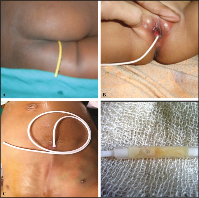
Results: The distal end of VP shunt was extruded from the anus (n = 3), vagina (n = 2), and anterior abdominal wall (n = 3). In five of these patients, shunt catheter was draining cerebrospinal fluid (CSF), the children were afebrile and CSF was sterile. In three children with extrusion of the shunt through the abdominal wall, the shunt tract was infected. Two of these patients had abscess in the shunt tract, which required incision and drainage. Both these patients had meningitis with a growth of Streptococcus species from CSF. Seven patients required further CSF diversion such as endoscopic third ventriculostomy (n = 3) or placement of VP shunt (n = 4).
Conclusion: Distal tip migration of VP shunt may prove to have potentially serious complications such as meningitis. A prompt and aggressive protocol of management is recommended.
Keywords: Abdominal end migration; extrusion; shunt complications; transabdominal; transanal; transvaginal.
Conflict of interest statement
There are no conflicts of interest.
Figures

References
-
- Ghritlaharey RK. Extrusion of ventriculoperitoneal shunt catheter through mouth in a two-year-old girl: a case report. Int J Clin Pediatr Surg. 2015;1:1–4.
-
- Griffith JA, DeFeo D. Peroral extrusion of a ventriculoperitoneal shunt catheter. Neurosurgery. 1987;21:259–61. - PubMed
-
- Mozingo JR, Cauthen JC. Vaginal perforation by a Raimondi peritoneal catheter in an adult. Surg Neurol. 1974;2:195–6. - PubMed
