Review
. 2018 Oct 1;9(5):924-937.
doi: 10.14336/AD.2017.1126. eCollection 2018 Oct.

Mitochondria in Ischemic Stroke: New Insight and Implications

Affiliations
Review

Mitochondria in Ischemic Stroke: New Insight and Implications

Fan Liu et al. Aging Dis. .

Abstract

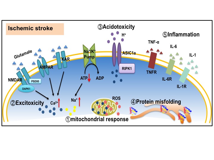
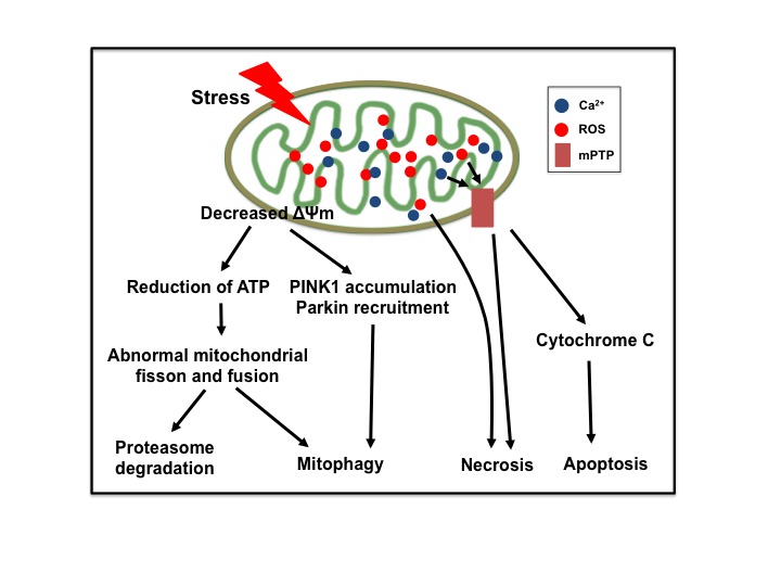
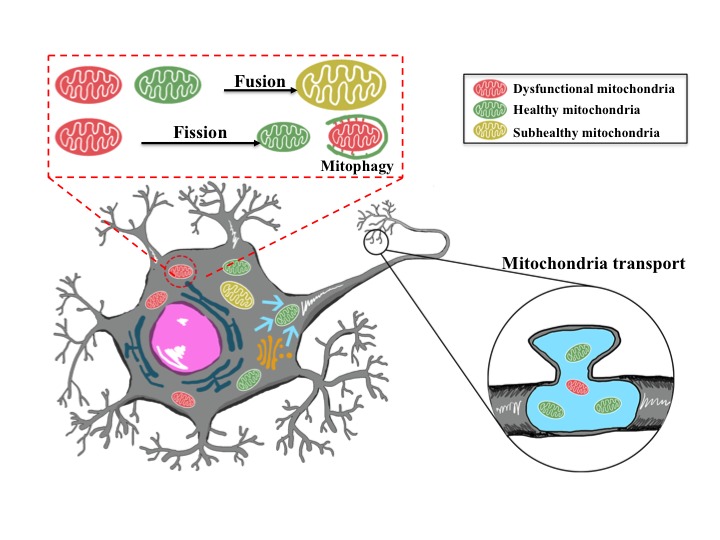
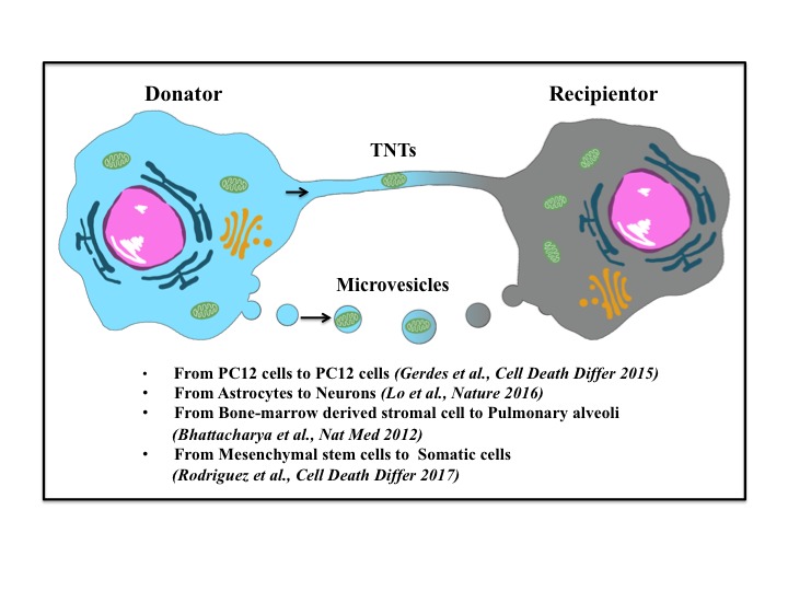
Stroke is the leading cause of death and adult disability worldwide. Mitochondrial dysfunction has been regarded as one of the hallmarks of ischemia/reperfusion (I/R) induced neuronal death. Maintaining the function of mitochondria is crucial in promoting neuron survival and neurological improvement. In this article, we review current progress regarding the roles of mitochondria in the pathological process of cerebral I/R injury. In particular, we emphasize on the most critical mechanisms responsible for mitochondrial quality control, as well as the recent findings on mitochondrial transfer in acute stroke. We highlight the potential of mitochondria as therapeutic targets for stroke treatment and provide valuable insights for clinical strategies.

Keywords: Ischemic stroke; mitochondrial quality control; mitochondrial transfer; mitophagy; neuroprotection.


Figures

Figure 1.
Figure 1.. Mechanisms underlying neuronal death in ischemic stroke
(1) Mitochondrial response, including excessive ROS production, mitochondrial calcium overloading, and disrupted mitochondria quality control. (2) Excitotoxicity. Excessive glutamate release and impeded reuptake of excitatory amino acids result in the activation of NMDARs, AMPARs and KARs. (3) Acidotoxity. Extracellular acidification leads to ischemic neuronal death by activating acid-sensing ion channel 1a (ASIC1a). (4) Protein misfolding. Protein misfolding and aggregation are observed after brain ischemia. (5) Inflammatory reaction. Microglia are activated and release cytokines and chemokines to induce inflammation reaction. All the factors mentioned above work synergistically to trigger cell death pathways such as apoptosis, necroptosis and autophagy. ROS: reactive oxygen species; AMPAR: α-amino-3-hydroxy-5-methyl-4-isoxazole-propionic acid receptor; NMDAR: N-methyl-D-aspartate receptor; KAR: kainite receptor; DAPK1: death associated protein kinase 1; PSD95: postsynaptic density protein 95; ASIC1a: acid-sensing ion channel 1a; RIPK1: receptor interacting protein kinase 1; TNF-α: tumor necrosis factor-α; IL-6: Interleukin 6; IL-1: Interleukin 1.
Figure 2.
Figure 2.. Mitochondria play a central role in ischemic neuronal death
Ischemia triggers the depolarization of mitochondrial membrane potential (ΔΨm), reduction of ATP production, accumulation of PINK1, recruitment of Parkin, overproduction of reactive oxygen species (ROS), overloading of matrix calcium, and opening of mitochondrial permeability transition pore (mPTP), eventually leading to neuronal death.
Figure 3.
Figure 3.. Mitochondrial fusion, fission or transport in neurons
Damaged mitochondria can be repaired through fusion with healthy mitochondria, and mitochondrial fission enables the segregation of damaged mitochondria and subsequent elimination via mitophagy. Mitochondria are transported and packed at axonal synapses, and are essential for neuronal transmission and plasticity.
Figure 4.
Figure 4.. Intercellular mitochondrial transfer
Mitochondria can be released by donate cells and uptake by recipient cells. Stressed or dying cells release mitochondria through tunneling nanotubes (TNTs) or microvesicles. Mitochondrial transfer can occur between same or different cell types.

References

    1. Qureshi AI, Tuhrim S, Broderick JP, Batjer HH, Hondo H, Hanley DF (2001). Spontaneous intracerebral hemorrhage. N Engl J Med, 344:1450-1460. - PubMed
    1. Lapchak PA, Zhang JH (2017). The High Cost of Stroke and Stroke Cytoprotection Research. Transl Stroke Res, 8:307-317. - PMC - PubMed
    1. Simao F, Ustunkaya T, Clermont AC, Feener EP (2017). Plasma kallikrein mediates brain hemorrhage and edema caused by tissue plasminogen activator therapy in mice after stroke. Blood, 129:2280-2290. - PMC - PubMed
    1. Adibhatla RM, Hatcher JF (2008). Tissue plasminogen activator (tPA) and matrix metalloproteinases in the pathogenesis of stroke: therapeutic strategies. CNS Neurol Disord Drug Targets, 7:243-253. - PMC - PubMed
    1. Henninger N, Fisher M (2016). Extending the Time Window for Endovascular and Pharmacological Reperfusion. Transl Stroke Res, 7:284-293. - PubMed

LinkOut - more resources