

Autologous and heterotopic transplantation of adipose stromal vascular fraction ameliorates stress urinary incontinence in rats with simulated childbirth trauma
- PMID: 30271860
- PMCID: PMC6147152
- DOI: 10.1016/j.reth.2017.11.003
Autologous and heterotopic transplantation of adipose stromal vascular fraction ameliorates stress urinary incontinence in rats with simulated childbirth trauma
Abstract
Introduction: Autologous transplantation of adipose stromal vascular fraction (SVF) is a cost-effective and technically accessible option for cell therapy. Clinical study of SVF transplantation for male stress urinary incontinence (SUI) is underway, but the effectiveness remains unknown for female SUI, majority of which is caused by childbirth trauma.
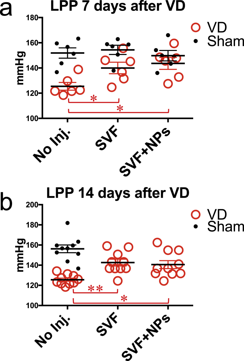
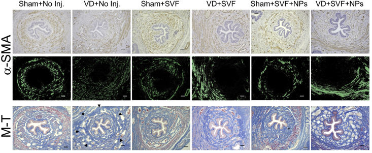
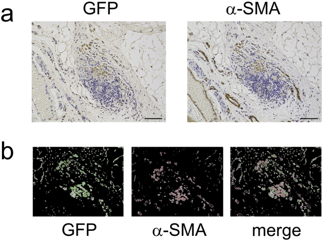
Methods: Vaginal Distension (VD) rats were generated as in vivo model for female SUI. To quantitate the severity of SUI, leak point pressure (LPP) was measured by placing a bladder catheter. There was a characteristic waveform of LPP with two-peaks, and we counted the second peak as an LPP value. Adipose SVF was separated from inguinal fat and delivered into external urethral sphincter (EUS) through transperineal injection. LPP was measured 7 or 14 days after SVF transplantation. Tissue damage and collagen synthesis around the EUS were visualized by Masson's trichrome and eosin staining. Antibody against α-smooth muscle actin (α-SMA) was used to stain smooth muscle or activated stromal cells. Donor SVF cells were distinguished from recipient EUS tissue by tracking with GFP transgene.
Results: VD procedure decreased the frequency at which the normal LPP waveform appeared and lowered the LPP value. SVF injection normalized the waveform as well as the level of LPP. VD disrupted histological structure of EUS and SVF failed to differentiate into striatal muscles. Instead, SVF increased α-SMA positive cells and collagen synthesis but the phenomena depended on VD stimulus. GFP tracking indicated that the transplanted SVF cells persisted for four weeks and synthesized α-SMA protein simultaneously.
Conclusions: Autologous transplantation of adipose SVF displayed bulking effects through collagen synthesis. However, such heterotopic activation was dependent on tissue damage.
Keywords: Adipose stromal vascular fraction; Collagen synthesis; EUS, external urethral sphincter; LPP, leak point pressure; Leak point pressure; NPs, low-molecular-weight heparin/protamine micro/nanoparticles; PNT, pudendal nerve transection; SUI, stress urinary incontinence:; SVF, stromal vascular fraction; Stress urinary incontinence; VD, vaginal distension; Vaginal distension model rat.
Figures


References
-
- Bourin P., Bunnell B.A., Casteilla L., Dominici M., Katz A.J., March K.L. Stromal cells from the adipose tissue-derived stromal vascular fraction and culture expanded adipose tissue-derived stromal/stem cells: a joint statement of the International Federation for Adipose Therapeutics and Science (IFATS) and the International Society for Cellular Therapy (ISCT) Cytotherapy. 2013;15:641–648. - PMC - PubMed
-
- Yamamoto T., Gotoh M., Hattori R., Toriyama K., Kamei Y., Iwaguro H. Periurethral injection of autologous adipose-derived stem cells for the treatment of stress urinary incontinence in patients undergoing radical prostatectomy: report of two initial cases. Int J Urol. 2010;17:75–82. - PubMed
-
- Yamamoto T., Gotoh M., Kato M., Majima T., Toriyama K., Kamei Y. Periurethral injection of autologous adipose-derived regenerative cells for the treatment of male stress urinary incontinence: report of three initial cases. Int J Urol. 2012;19:652–659. - PubMed
-
- Gotoh M., Yamamoto T., Kato M., Majima T., Toriyama K., Kamei Y. Regenerative treatment of male stress urinary incontinence by periurethral injection of autologous adipose-derived regenerative cells: 1-year outcomes in 11 patients. Int J Urol. 2014;21:294–300. - PubMed
LinkOut - more resources
Full Text Sources
