

Background-deflection Brillouin microscopy reveals altered biomechanics of intracellular stress granules by ALS protein FUS
- PMID: 30272018
- PMCID: PMC6131551
- DOI: 10.1038/s42003-018-0148-x
Background-deflection Brillouin microscopy reveals altered biomechanics of intracellular stress granules by ALS protein FUS
Abstract
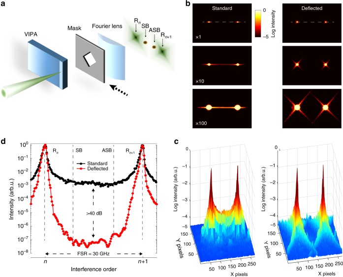
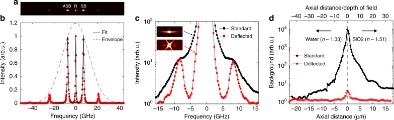
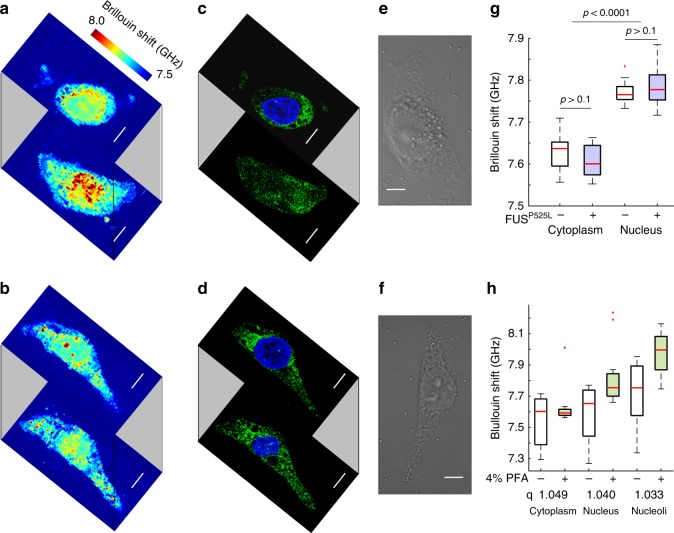
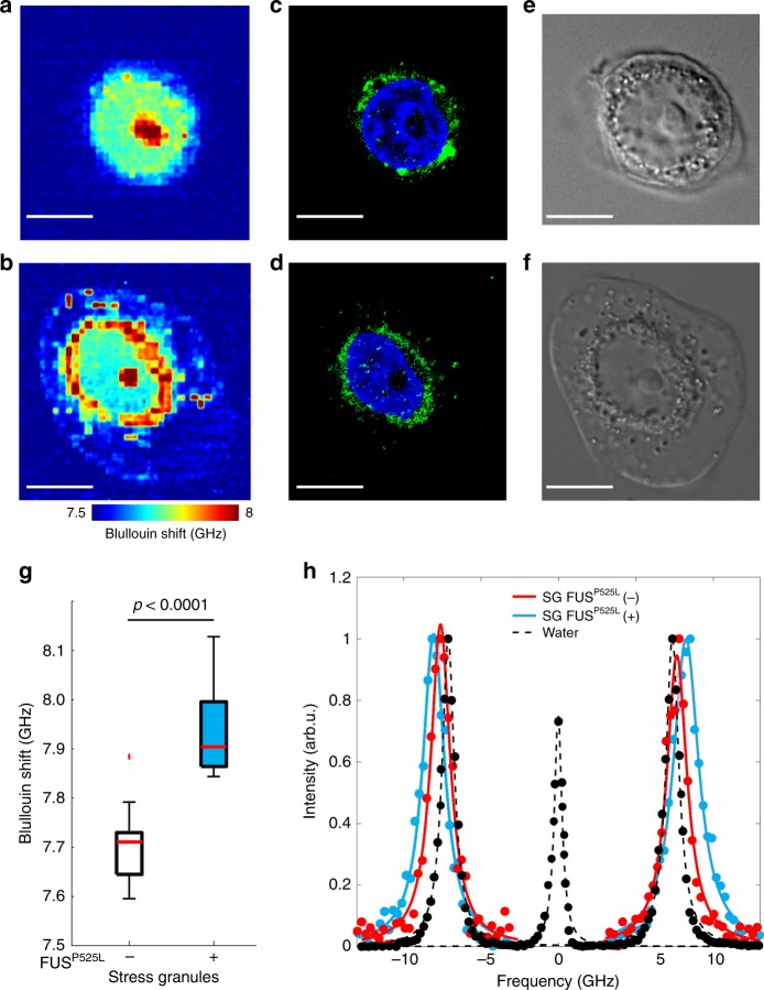
Altered cellular biomechanics have been implicated as key photogenic triggers in age-related diseases. An aberrant liquid-to-solid phase transition, observed in in vitro reconstituted droplets of FUS protein, has been recently proposed as a possible pathogenic mechanism for amyotrophic lateral sclerosis (ALS). Whether such transition occurs in cell environments is currently unknown as a consequence of the limited measuring capability of the existing techniques, which are invasive or lack of subcellular resolution. Here we developed a non-contact and label-free imaging method, named background-deflection Brillouin microscopy, to investigate the three-dimensional intracellular biomechanics at a sub-micron resolution. Our method exploits diffraction to achieve an unprecedented 10,000-fold enhancement in the spectral contrast of single-stage spectrometers, enabling, to the best of our knowledge, the first direct biomechanical analysis on intracellular stress granules containing ALS mutant FUS protein in fixed cells. Our findings provide fundamental insights on the critical aggregation step underlying the neurodegenerative ALS disease.
Conflict of interest statement
The authors declare no competing interests.
Figures
Similar articles
-
ALS-causing hPFN1 mutants differentially disrupt LLPS of FUS prion-like domain.Biochem Biophys Res Commun. 2023 Jul 5;664:35-42. doi: 10.1016/j.bbrc.2023.04.101. Epub 2023 Apr 28. Biochem Biophys Res Commun. 2023. PMID: 37130459
-
ALS-Related Mutant FUS Protein Is Mislocalized to Cytoplasm and Is Recruited into Stress Granules of Fibroblasts from Asymptomatic FUS P525L Mutation Carriers.Neurodegener Dis. 2017;17(6):292-303. doi: 10.1159/000480085. Epub 2017 Oct 17. Neurodegener Dis. 2017. PMID: 29035885
-
Mechanism underlying liquid-to-solid phase transition in fused in sarcoma liquid droplets.Phys Chem Chem Phys. 2022 Aug 17;24(32):19346-19353. doi: 10.1039/d2cp02171d. Phys Chem Chem Phys. 2022. PMID: 35943083
-
Amyotrophic Lateral Sclerosis, FUS and Protein Synthesis Defects.Stem Cell Rev Rep. 2023 Apr;19(3):625-638. doi: 10.1007/s12015-022-10489-8. Epub 2022 Dec 14. Stem Cell Rev Rep. 2023. PMID: 36515764 Review.
-
Pathophysiological implications of RNP granules in frontotemporal dementia and ALS.Neurochem Int. 2020 Nov;140:104819. doi: 10.1016/j.neuint.2020.104819. Epub 2020 Aug 5. Neurochem Int. 2020. PMID: 32763254 Review.
Cited by
-
Mutant FUS and ELAVL4 (HuD) Aberrant Crosstalk in Amyotrophic Lateral Sclerosis.Cell Rep. 2019 Jun 25;27(13):3818-3831.e5. doi: 10.1016/j.celrep.2019.05.085. Cell Rep. 2019. PMID: 31242416 Free PMC article.
-
Quantifying cellular forces and biomechanical properties by correlative micropillar traction force and Brillouin microscopy.Biomed Opt Express. 2019 Apr 3;10(5):2202-2212. doi: 10.1364/BOE.10.002202. eCollection 2019 May 1. Biomed Opt Express. 2019. PMID: 31149370 Free PMC article.
-
Multipass etalon cascade for high-resolution parallel spectroscopy.Opt Lett. 2021 Feb 15;46(4):781-784. doi: 10.1364/OL.418090. Opt Lett. 2021. PMID: 33577513 Free PMC article.
-
The mechanobiology of biomolecular condensates.Biophys Rev (Melville). 2025 Mar 25;6(1):011310. doi: 10.1063/5.0236610. eCollection 2025 Mar. Biophys Rev (Melville). 2025. PMID: 40160200 Free PMC article. Review.
-
Probing molecular crowding in compressed tissues with Brillouin light scattering.Proc Natl Acad Sci U S A. 2022 Jan 25;119(4):e2113614119. doi: 10.1073/pnas.2113614119. Proc Natl Acad Sci U S A. 2022. PMID: 35046032 Free PMC article.
References
LinkOut - more resources
Full Text Sources
Other Literature Sources
Miscellaneous
