

A new histological evaluation method to detect residual ophthalmic viscosurgical devices for cataract surgery
- PMID: 30272034
- PMCID: PMC6159335
- DOI: 10.1016/j.heliyon.2018.e00822
A new histological evaluation method to detect residual ophthalmic viscosurgical devices for cataract surgery
Abstract
Purpose: To establish a new evaluation method to quantify residual ophthalmic viscosurgical device (OVD) volume and corneal endothelium adhesion properties for phacoemulsification surgery.
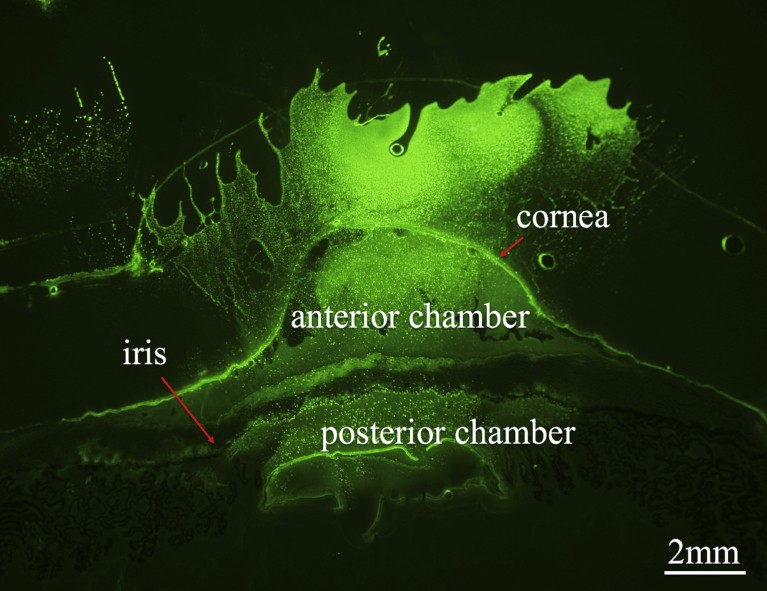
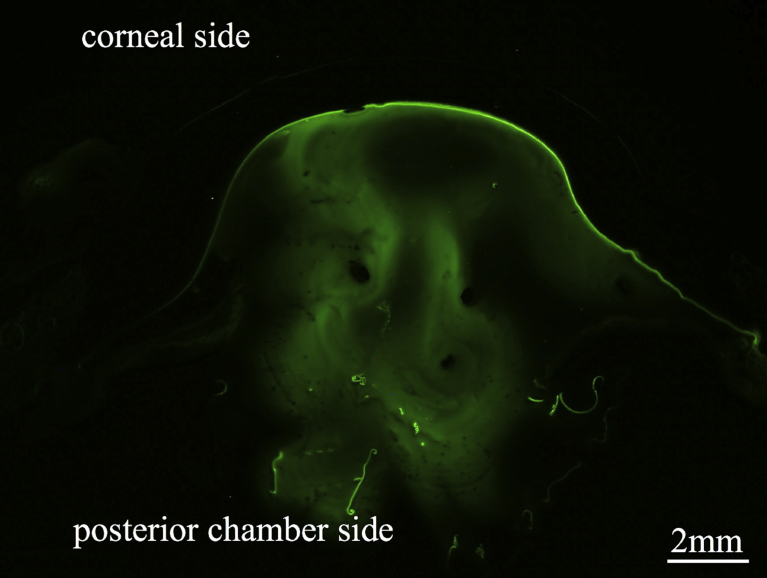
Methods: We compared the performance of four OVDs (Viscoat®, Healon5®, Healon® and DisCoVisc®) using porcine eyes. First, OVDs were mixed with fluorescent-conjugated dextrans to render them visible under the microscope. A corneal side port was opened, followed by a continuous curvilinear capsulorhexis, and a corneal tunnel incision was made. OVDs were injected, then the lens was removed using one-handed phacoemulsification. After this procedure, the anterior segment of the eye was isolated via an equatorial incision and the tissue was immediately frozen in shimmering liquid nitrogen. Sagittal slices (20 μm) were cut with a Cryostat from limbus to limbus. Every tenth slide was imaged using a fluorescent microscope with a CCD camera. We evaluated the percentage of the corneal endothelium covered by each OVD as the OVD adhesion to corneal endothelium ratio (OAE ratio) and the volume of residual OVD in the anterior chamber.
Results: Viscoat® showed significantly higher endothelium coverage compared with both Healon® and DisCoVisc®. A statistically larger volume of Healon5® remained in the anterior chamber compared with Healon® and DisCoVisc®.
Conclusion: The new evaluation methods used here provide precise quantitative analysis of OAE ratio and residual OVD volume. These results show that Viscoat® and Healon5® have a high potential for coating the corneal endothelium during phacoemulsification and aspiration surgery.
Keywords: Ophthalmology; Surgery.
Figures


Similar articles
-
Thickness of the Protective Layers of Different Ophthalmic Viscosurgical Devices During Lens Surgery in a Porcine Model.Transl Vis Sci Technol. 2022 Feb 1;11(2):28. doi: 10.1167/tvst.11.2.28. Transl Vis Sci Technol. 2022. PMID: 35175318 Free PMC article.
-
Quantitative assessment of ophthalmic viscosurgical device retention using in vivo confocal microscopy.J Cataract Refract Surg. 2005 Dec;31(12):2363-8. doi: 10.1016/j.jcrs.2005.05.032. J Cataract Refract Surg. 2005. PMID: 16473232
-
Retention and removal of a new viscous dispersive ophthalmic viscosurgical device during cataract surgery in animal eyes.Br J Ophthalmol. 2006 Apr;90(4):485-7. doi: 10.1136/bjo.2005.085969. Br J Ophthalmol. 2006. PMID: 16547332 Free PMC article.
-
Corneal Outcomes Following Cataract Surgery Using Ophthalmic Viscosurgical Devices Composed of Chondroitin Sulfate-Hyaluronic Acid: A Systematic Review and Meta-Analysis.Clin Ophthalmol. 2023 Jul 24;17:2083-2096. doi: 10.2147/OPTH.S419863. eCollection 2023. Clin Ophthalmol. 2023. PMID: 37521151 Free PMC article. Review.
-
Free radical development in phacoemulsification cataract surgery.J Nippon Med Sch. 2005 Feb;72(1):4-12. doi: 10.1272/jnms.72.4. J Nippon Med Sch. 2005. PMID: 15834202 Review.
Cited by
-
Thickness of the Protective Layers of Different Ophthalmic Viscosurgical Devices During Lens Surgery in a Porcine Model.Transl Vis Sci Technol. 2022 Feb 1;11(2):28. doi: 10.1167/tvst.11.2.28. Transl Vis Sci Technol. 2022. PMID: 35175318 Free PMC article.
References
-
- Kelman C.D. Phaco-emulsification and aspiration: a new techniques of cataract removal. Am. J. Ophthalmol. 1967;64:23–35. PMID: 6028631. - PubMed
-
- McCarey B.E., Polack F.M., Marshall W. The phacoemulsification procedure. I. The effect of intraocular irrigating solutions on the corneal endothelium. Investig. Ophthalmol. 1976;15:449–457. PMID: 931689. - PubMed
-
- Polack F.M., Sugar A. The phacoemulsification procedure. II. Corneal endothelial changes. Investig. Ophthalmol. 1976;15:458–469. PMID: 931690. - PubMed
-
- Binder P.S., Sternberg H., Wickman M.G., Worthen D.M. Corneal endothelial damage with phacoemulsification. Am. J. Ophthalmol. 1976;82:48–54. PMID: 937457. - PubMed
-
- Irvine A.R., Kratz R.P., O’Donnel J.J. Endothelial damage with phacoemulsification and intraocular lens implantation. Arch. Ophthalmol. 1978;96:1023–1026. PMID: 655939. - PubMed
LinkOut - more resources
Full Text Sources
