

Early Developmental Trajectories of Functional Connectivity Along the Visual Pathways in Rhesus Monkeys
- PMID: 30272135
- PMCID: PMC6644858
- DOI: 10.1093/cercor/bhy222
Early Developmental Trajectories of Functional Connectivity Along the Visual Pathways in Rhesus Monkeys
Abstract
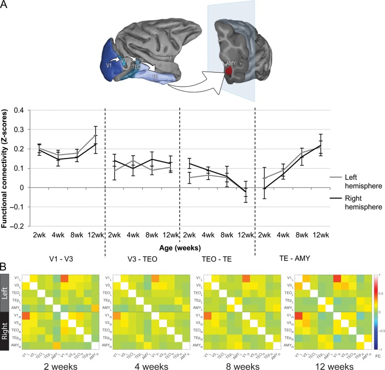
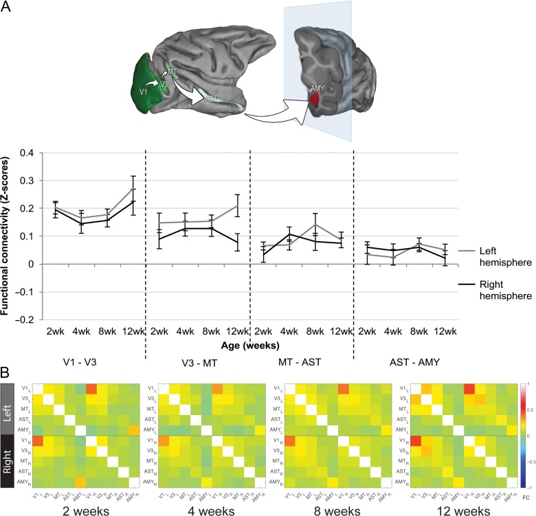
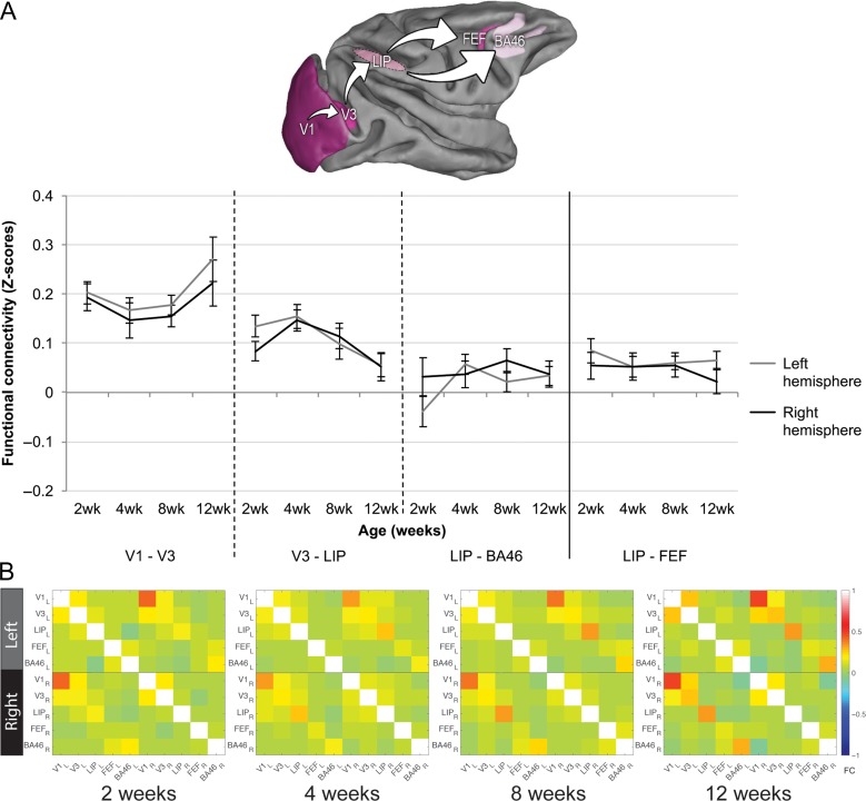
Early social interactions shape the development of social behavior, although the critical periods or the underlying neurodevelopmental processes are not completely understood. Here, we studied the developmental changes in neural pathways underlying visual social engagement in the translational rhesus monkey model. Changes in functional connectivity (FC) along the ventral object and motion pathways and the dorsal attention/visuo-spatial pathways were studied longitudinally using resting-state functional MRI in infant rhesus monkeys, from birth through early weaning (3 months), given the socioemotional changes experienced during this period. Our results revealed that (1) maturation along the visual pathways proceeds in a caudo-rostral progression with primary visual areas (V1-V3) showing strong FC as early as 2 weeks of age, whereas higher-order visual and attentional areas (e.g., MT-AST, LIP-FEF) show weak FC; (2) functional changes were pathway-specific (e.g., robust FC increases detected in the most anterior aspect of the object pathway (TE-AMY), but FC remained weak in the other pathways (e.g., AST-AMY)); (3) FC matures similarly in both right and left hemispheres. Our findings suggest that visual pathways in infant macaques undergo selective remodeling during the first 3 months of life, likely regulated by early social interactions and supporting the transition to independence from the mother.
Keywords: macaque; neurodevelopment; resting state functional MRI; social visual engagement.
© The Author(s) 2018. Published by Oxford University Press. All rights reserved. For Permissions, please e-mail: journals.permissions@oup.com.
Figures
Similar articles
-
The Development of Socially Directed Attention: A Functional Magnetic Resonance Imaging Study in Infant Monkeys.J Cogn Neurosci. 2024 Dec 1;36(12):2742-2760. doi: 10.1162/jocn_a_02187. J Cogn Neurosci. 2024. PMID: 38739568 Free PMC article.
-
Functional maturation in visual pathways predicts attention to the eyes in infant rhesus macaques: Effects of social status.Dev Cogn Neurosci. 2023 Apr;60:101213. doi: 10.1016/j.dcn.2023.101213. Epub 2023 Feb 8. Dev Cogn Neurosci. 2023. PMID: 36774827 Free PMC article.
-
Developmental outcomes of early adverse care on amygdala functional connectivity in nonhuman primates.Dev Psychopathol. 2020 Dec;32(5):1579-1596. doi: 10.1017/S0954579420001133. Dev Psychopathol. 2020. PMID: 33427167 Free PMC article.
-
Resting-state functional connectivity in individuals with bipolar disorder during clinical remission: a systematic review.J Psychiatry Neurosci. 2018 Aug;43(5):298-316. doi: 10.1503/jpn.170175. J Psychiatry Neurosci. 2018. PMID: 30125243 Free PMC article.
-
Neurodevelopment of the visual system in typically developing children.Prog Brain Res. 2011;189:113-36. doi: 10.1016/B978-0-444-53884-0.00021-X. Prog Brain Res. 2011. PMID: 21489386 Review.
Cited by
-
Use of connectotyping on task functional MRI data reveals dynamic network level cross talking during task performance.Front Neurosci. 2022 Oct 10;16:951907. doi: 10.3389/fnins.2022.951907. eCollection 2022. Front Neurosci. 2022. PMID: 36300171 Free PMC article.
-
Early developmental changes in visual social engagement in infant rhesus monkeys.Dev Cogn Neurosci. 2020 Jun;43:100778. doi: 10.1016/j.dcn.2020.100778. Epub 2020 Apr 18. Dev Cogn Neurosci. 2020. PMID: 32510341 Free PMC article.
-
Structural development of cortical lobes during the first 6 months of life in infant macaques.Dev Cogn Neurosci. 2021 Apr;48:100906. doi: 10.1016/j.dcn.2020.100906. Epub 2021 Jan 8. Dev Cogn Neurosci. 2021. PMID: 33465553 Free PMC article.
-
The effects of chronic social stress on cognitive flexibility in adult female macaques.bioRxiv [Preprint]. 2025 Aug 1:2025.08.01.667938. doi: 10.1101/2025.08.01.667938. bioRxiv. 2025. PMID: 40766392 Free PMC article. Preprint.
-
Lateralized Connectivity between Globus Pallidus and Motor Cortex is Associated with Freezing of Gait in Parkinson's Disease.Neuroscience. 2020 Sep 1;443:44-58. doi: 10.1016/j.neuroscience.2020.06.036. Epub 2020 Jul 3. Neuroscience. 2020. PMID: 32629155 Free PMC article.
References
-
- Andersson JL, Skare S, Ashburner J. 2003. How to correct susceptibility distortions in spin-echo echo-planar images: application to diffusion tensor imaging. Neuroimage. 20:870–888. - PubMed
-
- Bachevalier J, Hagger C, Mishkin M. 1991. Functional maturation of the occipitotemporal pathway in infant rhesus monkeys. In: Alfred Benzon Symposium No. 31: Brain Work and Brain Function, Munksgaard, Copenhagen, p. 231–240.
MeSH terms
Grants and funding
LinkOut - more resources
Full Text Sources
