

The Neuroanatomy of Autism Spectrum Disorder Symptomatology in 22q11.2 Deletion Syndrome
- PMID: 30272146
- PMCID: PMC6644859
- DOI: 10.1093/cercor/bhy239
The Neuroanatomy of Autism Spectrum Disorder Symptomatology in 22q11.2 Deletion Syndrome
Abstract
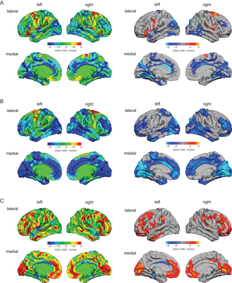
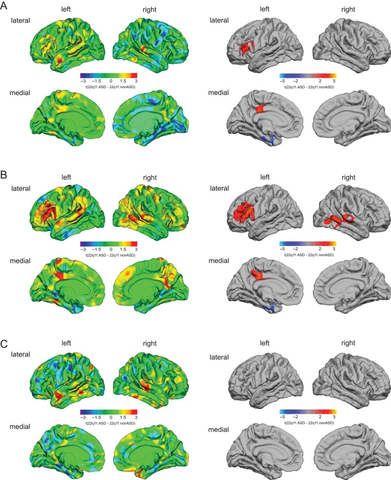
22q11.2 Deletion Syndrome (22q11.2DS) is a genetic condition associated with a high prevalence of neuropsychiatric conditions that include autism spectrum disorder (ASD). While evidence suggests that clinical phenotypes represent distinct neurodevelopmental outcomes, it remains unknown whether this translates to the level of neurobiology. To fractionate the 22q11.2DS phenotype on the level of neuroanatomy, we examined differences in vertex-wise estimates of cortical volume, surface area, and cortical thickness between 1) individuals with 22q11.2DS (n = 62) and neurotypical controls (n = 57) and 2) 22q11.2DS individuals with ASD symptomatology (n = 30) and those without (n = 25). We firstly observed significant differences in surface anatomy between 22q11.2DS individuals and controls for all 3 neuroanatomical features, predominantly in parietotemporal regions, cingulate and dorsolateral prefrontal cortices. We also established that 22q11.2DS individuals with ASD symptomatology were neuroanatomically distinct from 22q11.2DS individuals without ASD symptoms, particularly in brain regions that have previously been linked to ASD (e.g., dorsolateral prefrontal cortices and the entorhinal cortex). Our findings indicate that different clinical 22q11.2DS phenotypes, including those with ASD symptomatology, may represent different neurobiological subgroups. The spatially distributed patterns of neuroanatomical differences associated with ASD symptomatology in 22q11.2DS may thus provide useful information for patient stratification and the prediction of clinical outcomes.
Keywords: 22q11.2 Deletion Syndrome; autism spectrum disorder; brain anatomy; neurodevelopment; surface based morphometry.
© The Author(s) 2018. Published by Oxford University Press. All rights reserved. For Permissions, please e-mail: journals.permissions@oup.com.
Figures
Similar articles
-
Neuroanatomical underpinnings of autism symptomatology in carriers and non-carriers of the 22q11.2 microdeletion.Mol Autism. 2020 Jun 8;11(1):46. doi: 10.1186/s13229-020-00356-z. Mol Autism. 2020. PMID: 32513259 Free PMC article.
-
Patterns of Cortical Folding Associated with Autistic Symptoms in Carriers and Noncarriers of the 22q11.2 Microdeletion.Cereb Cortex. 2020 Sep 3;30(10):5281-5292. doi: 10.1093/cercor/bhaa108. Cereb Cortex. 2020. PMID: 32420595 Free PMC article.
-
22q11.2 duplication syndrome: elevated rate of autism spectrum disorder and need for medical screening.Mol Autism. 2016 May 6;7:27. doi: 10.1186/s13229-016-0090-z. eCollection 2016. Mol Autism. 2016. PMID: 27158440 Free PMC article.
-
Developmental trajectories in 22q11.2 deletion.Am J Med Genet C Semin Med Genet. 2015 Jun;169(2):172-81. doi: 10.1002/ajmg.c.31435. Epub 2015 May 18. Am J Med Genet C Semin Med Genet. 2015. PMID: 25989227 Free PMC article. Review.
-
[Neurocognitive and psychiatric management of the 22q11.2 deletion syndrome].Encephale. 2015 Jun;41(3):266-73. doi: 10.1016/j.encep.2014.10.005. Epub 2014 Dec 16. Encephale. 2015. PMID: 25523123 Review. French.
Cited by
-
Brain morphometry in 22q11.2 deletion syndrome: an exploration of differences in cortical thickness, surface area, and their contribution to cortical volume.Sci Rep. 2020 Nov 2;10(1):18845. doi: 10.1038/s41598-020-75811-1. Sci Rep. 2020. PMID: 33139857 Free PMC article.
-
Neuroanatomical underpinnings of autism symptomatology in carriers and non-carriers of the 22q11.2 microdeletion.Mol Autism. 2020 Jun 8;11(1):46. doi: 10.1186/s13229-020-00356-z. Mol Autism. 2020. PMID: 32513259 Free PMC article.
-
Neurobiological perspective of 22q11.2 deletion syndrome.Lancet Psychiatry. 2019 Nov;6(11):951-960. doi: 10.1016/S2215-0366(19)30076-8. Epub 2019 Aug 5. Lancet Psychiatry. 2019. PMID: 31395526 Free PMC article. Review.
-
Neuroimaging Phenotypes Associated With Risk and Resilience for Psychosis and Autism Spectrum Disorders in 22q11.2 Microdeletion Syndrome.Biol Psychiatry Cogn Neurosci Neuroimaging. 2021 Feb;6(2):211-224. doi: 10.1016/j.bpsc.2020.08.015. Epub 2020 Sep 6. Biol Psychiatry Cogn Neurosci Neuroimaging. 2021. PMID: 33218931 Free PMC article. Review.
-
Patterns of Cortical Folding Associated with Autistic Symptoms in Carriers and Noncarriers of the 22q11.2 Microdeletion.Cereb Cortex. 2020 Sep 3;30(10):5281-5292. doi: 10.1093/cercor/bhaa108. Cereb Cortex. 2020. PMID: 32420595 Free PMC article.
References
-
- Amaral DG, Schumann CM, Nordahl CW. 2008. Neuroanatomy of autism. Trends Neurosci. 31(3):137–145. - PubMed
-
- Antshel KM, AbdulSabur N, Roizen N, Fremont W, Kates WR. 2005. Sex differences in cognitive functioning in velocardiofacial syndrome (VCFS). Dev Neuropsychol. 28(3):849–869. - PubMed
-
- Antshel KM, Aneja A, Strunge L, Peebles J, Fremont WP, Stallone K, AbdulSabur N, Higgins AM, Shprintzen RJ, Kates WR. 2007. Autistic spectrum disorders in velo-cardio facial syndrome (22q11.2 deletion). J Autism Dev Disord. 37(9):1776–1786. - PubMed
-
- Blakemore SJ. 2008. The social brain in adolescence. Nat Rev Neurosci. 9(4):267–277. - PubMed
-
- Castelli F, Frith C, Happe F, Frith U. 2002. Autism, asperger syndrome and brain mechanisms for the attribution of mental states to animated shapes. Brain. 125:1839–1849. - PubMed
