. 2018 Dec 14;46(22):11726-11742.
doi: 10.1093/nar/gky871.

Unusual dicistronic expression from closely spaced initiation codons in an umbravirus subgenomic RNA

Affiliations

Unusual dicistronic expression from closely spaced initiation codons in an umbravirus subgenomic RNA

Feng Gao et al. Nucleic Acids Res. .

Abstract

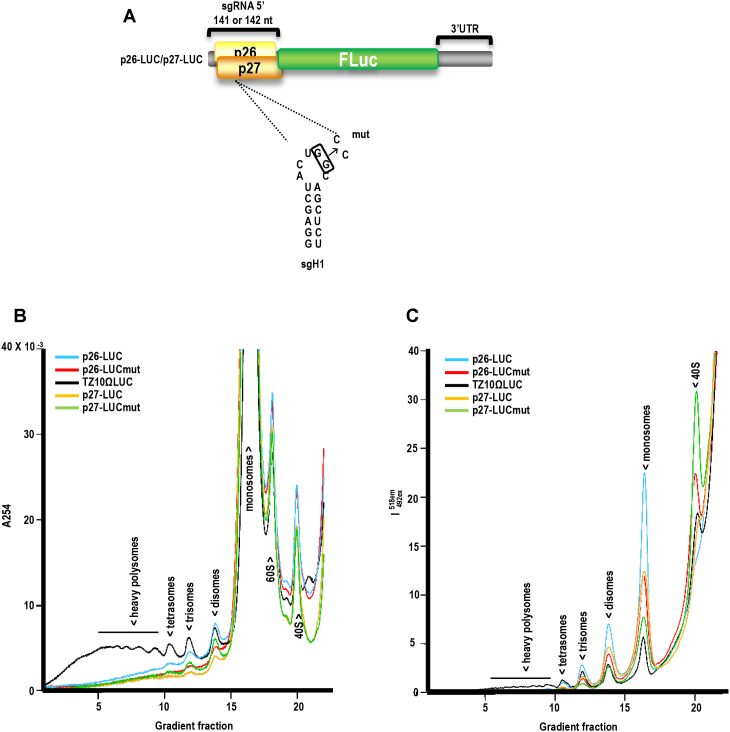
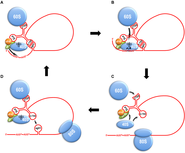
Translation commencing at closely spaced initiation codons is common in RNA viruses with limited genome space. In the subgenomic RNA (sgRNA) of Pea enation mosaic virus 2, two closely spaced, out-of-frame start codons direct synthesis of movement/stability proteins p26 and p27. Efficient translation from AUG26/AUG27 is dependent on three 3'-proximal cap-independent translation enhancers (3'CITEs), whereas translation of the genomic (gRNA) requires only two. Contrary to strictly scanning-dependent initiation at the gRNA, sequence context of AUG26/AUG27 does not conform with Kozak requirements and insertion of efficient upstream AUGs had pronounced effects for AUG26 but only moderate effects for AUG27. Insertion of a hairpin within an extended 5' UTR did not significantly impact translation from AUG26/AUG27. Furthermore, AUG27 repressed translation from upstream AUG26 and this effect was mitigated when inter-codon spacing was reduced. Addition of a stable hairpin to the very 5' end of the sgRNA severely restricted translation, testifying that this 3'CITE-driven initiation is 5' end-dependent. Similar to gRNA, sgRNA reporter transcripts were nearly exclusively associated with light polysomes and 3'CITE-promoted long-distance interaction connecting the sgRNA ends affected the number of templates translated and not the initiation rate. We propose a non-canonical, 3'CITE-driven mechanism for efficient dicistronic expression from umbravirus sgRNAs.


Figures

Figure 1.
Figure 1.
5′ and 3′ sequences involved in translation of the PEMV2 sgRNA. (A) Genome organization of PEMV2 gRNA and sgRNA. (B) Sequences at the 5′ end 3′ ends of the sgRNA. Initiation codons for AUG26 and AUG27 are in blue and red, respectively. The long-distance interaction (LDI) is shown in green. The kl-TSS 3′CITE binds to 40S, 60S and 80S ribosomes and can simultaneously engage in the LDI (34,41). The PTE 3′CITE binds to eIF4E, which is followed by binding to eIF4G (35). The TSS 3′CITE binds to 60S and 80S ribosomes (42).
Figure 2.
Figure 2.
Effect of upstream AUG codons on translation initiation. (A) Alterations to the gRNA 5′ UTR in a luciferase reporter construct. U7G generates an out-of-frame AUG upstream of the natural AUG at position 21. (B) In vivo translation of WT and mutant reporter constructs in Arabidopsis thaliana protoplasts. Throughout this report, mean values and standard error were calculated from at least three independent experiments. One-way ANOVA was used to analyze the statistical significance; *P ≤ 0.05, **P ≤ 0.01. (C) Sequences of experimental and control full-length sgRNA constructs. Inserted and altered sequences are in orange. New initiation codons are in green. Additional altered residues in +37AUG4 to improve the context of the inserted AUG are in red. (D) Translation of WT and +37 sgRNA transcripts in WGE. (E) Translation of WT and +37 mutant reporter constructs in Arabidopsis protoplasts. (F) Typical SDS-PAGE gel showing levels of p26 and p27, and new p26 extended products (p26*) generated from the added in-frame initiation codons following translation in WGE. p26 and p27 migrate aberrantly, as previously described (37). (G) Quantification of the relative levels of p26*, p26 and p27 of +37 derived constructs in WGE. p26 and p27 protein levels were normalized to 100 for ease in comparisons. p26* levels were normalized to p26 for comparison. (H) (left) Typical SDS-PAGE gel showing levels of p26, p27 and the new product generated from the downstream, in-frame AUG (p27*); (right), quantification of the relative levels of p26 and p27 in WGE.
Figure 3.
Figure 3.
Effect of upstream stable hairpin on translation from AUG26 and AUG27. (A) Forty nucleotide hairpin (HP) generated from the 37 nt added sequence. (B) Constructs showing insertion of HP at the 5′ end of WT sgRNA (5′HP), at the 5′ end prior to a 100-nt insert (+137–5′HP); and in the center of the 100-nt insert (+137-cHP). Insertion of unstructured sequence at the 5′ end prior to a 100-nt insert serves as a control (+137). (C) In vitro translation of wt and mutant sgRNAs. Data were either normalized to p26 and p27 levels in WT (left) or +137 (right). (D) Translation of p26-LUC and p27-LUC in protoplasts containing the 5′ terminal HP or 5′ terminal extensions (left) or +137cHP (right). One-way ANOVA was used to analyze the statistical significance; *P ≤ 0.05, **P ≤ 0.01. See legend to Figure 2 for more details.
Figure 4.
Figure 4.
Effect of context on translation initiation from AUG26 and AUG27. (A) Mutations in and near AUG26 and AUG27 that either alter the AUGs (m1 and m2) or improve their Kozak context (m3 and m4) are shown for WT constructs and constructs with added 5′ +37 nt. Data for m1 and m2 are from reference (37) and are repeated here for clarity. (B) Translation of WT and mutant sgRNAs in WGE. (C) Relative luciferase activity in protoplasts transfected with WT and mutant p26-LUC and p27-LUC. (D) Translation of +37 and +37 mutant sgRNAs in WGE. (E) Translation of WT, +37 and +37 mutant reporter constructs in Arabidopsis protoplasts. B, C, D, and E, one-way ANOVA was used to analyze the statistical significance; *P ≤ 0.05, **P ≤ 0.01.See legend to Figure 2 for more details. Luciferase activity that persists in vivo despite the lack of initiation codons in +37m1 and +37m2 is likely due to translation initiating from the natural luciferase AUG. (F) Ribosome toeprinting of WT and mutant using WGE. Toeprints corresponding to stalled ribosomes on AUG26 and AUG27 are labeled. Single and double asterisks denote additional AUG27 and AUG26 toeprints, respectively, described previously (37), which may represent improper fixing of the sgRNA in the mRNA-binding cleft of the 40S subunit, allowing for further extension by the reverse transcriptase (33). G, U, C, A are ladder lanes. Data are from three independent experiments. Line between lanes denotes removal of an intervening lane.
Figure 5.
Figure 5.
Replacing the 5′ UTR of PEMV2 sgRNA with the 5′ UTRs of other umbraviruses. (A) Sequences used to replace the 5′ UTR of PEMV2 sgRNA. TBTV+1 has an insert of a single residue (in orange) such that the sequence is the same length as the 5′ UTRs of PEMV2 and OPMV, and now differs at a single position from the OPMV 5′ UTR sequence. (B) In vitro translation of WT and mutant sgRNAs in WGE. (C) In vivo translation of WT and mutant p26-LUC and p27-LUC in protoplasts. B and C, one-way ANOVA was used to analyze the statistical significance; *P ≤ 0.05, **P ≤ 0.01. (D) Ribosome toeprinting of WT and mutant sgRNAs in WGE. Single and double asterisks denote additional AUG27 and AUG26 toeprints, respectively.
Figure 6.
Figure 6.
Effect of shortening and lengthening the distance between AUG26 and AUG27 on start codon selection. (A) Constructs showing deletions and insertions between AUG26 and AUG27, and deletions between AUG27 and sgH1. (B) In vitro translation of WT and mutant sgRNAs in WGE. (C) In vivo translation of WT and mutant p26-LUC and p27-LUC in protoplasts. One-way ANOVA was used to analyze the statistical significance; *P ≤ 0.05, **P ≤ 0.01. See legend to Figure 2 for more details.
Figure 7.
Figure 7.
LDI enhances the number of templates translated but not the number of ribosomes per template. (A) Luciferase constructs p26-LUC and p27-LUC showing mutation in sgH1 that eliminates the LDI between the kl-TSS and sgH1, generating p26-LUCmut and p27-LUCmut. (B) Polysome distribution on uncapped p26-LUC, p26-LUCmut, p27-LUC, p27-LUCmut and TZ10ΩLuc templates. TZ10ΩLuc is a control template containing the TMV Ω translation enhancer in the 5′ UTR and the TMV 3′ UTR. RNAs (50 nM) were incubated in WGE for 30 min, then subjected to sedimentation analysis. Ultraviolet absorbance profiles reflect mainly the distribution of ribosomes. (C) Fluorescence profiles represent allocation of the labeled reporter RNAs along gradients.
Figure 8.
Figure 8.
Model for translation of PEMV2 sgRNA. (A) Long-distance RNA–RNA interaction provides the delivery of 3′CITE-bound ribosomal subunits and initiation factors to 5′-termini of the sgRNA to engage the pre-formed initiation complex in recognition of the RNA 5′-end and further 5′ UTR scanning. (B) kl-TSS/sgH1 kissing-loop interaction allocates scanning 40S subunit at certain sterically favorable position along the RNA chain and engages it in transient sampling of nearby AUG26 and AUG27, with some preference for the latter. Following stable closed complex formation at one of the AUGs, the 40S subunit is joined by TSS-bound 60S. (C) Newly formed 80S ribosome starts translation of a reading frame and promptly melts the sgH1 stem-loop, tearing sgRNA ends apart and suspending the initiation process. Meanwhile, 3′CITEs are reloaded with new ribosomal subunits. (D) After the 80S has passed, sgH1 structure is restored again allowing inter-termini interaction to promote a new initiation cycle.

Similar articles

Cited by

References

    1. Jackson R.J., Hellen C.U., Pestova T.V.. The mechanism of eukaryotic translation initiation and principles of its regulation. Nat. Rev. Mol. Cell Biol. 2010; 11:113–127. - PMC - PubMed
    1. Kozak M. Initiation of translation in prokaryotes and eukaryotes. Gene. 1999; 234:187–208. - PubMed
    1. Gualerzi C.O., Pon C.L.. Initiation of mRNA translation in prokaryotes. Biochemistry. 1990; 29:5881–5889. - PubMed
    1. Sonenberg N., Hinnebusch A.G.. Regulation of translation initiation in eukaryotes: mechanisms and biological targets. Cell. 2009; 136:731–745. - PMC - PubMed
    1. Kozak M. The scanning model for translation: an update. J. Cell Biol. 1989; 108:229–241. - PMC - PubMed

Publication types