. 2019 Jan 1;80(1):94-102.
doi: 10.1097/QAI.0000000000001871.

Placental Structure in Preterm Birth Among HIV-Positive Versus HIV-Negative Women in Kenya

Affiliations

Placental Structure in Preterm Birth Among HIV-Positive Versus HIV-Negative Women in Kenya

Moses M Obimbo et al. J Acquir Immune Defic Syndr. .

Abstract

Background: Preterm birth (PTB) is a major cause of infant morbidity and mortality in developing countries. Recent data suggest that in addition to Human Immunodeficiency Virus (HIV) infection, use of antiretroviral therapy (ART) increases the risk of PTB. As the mechanisms remain unexplored, we conducted this study to determine whether HIV and ART were associated with placental changes that could contribute to PTB.

Setting: We collected and evaluated placentas from 38 HIV-positive women on ART and 43 HIV-negative women who had preterm deliveries in Nairobi, Kenya.

Methods: Anatomical features of the placentas were examined at gross and microscopic levels. Cases were matched for gestational age and compared by the investigators who were blinded to maternal HIV serostatus.

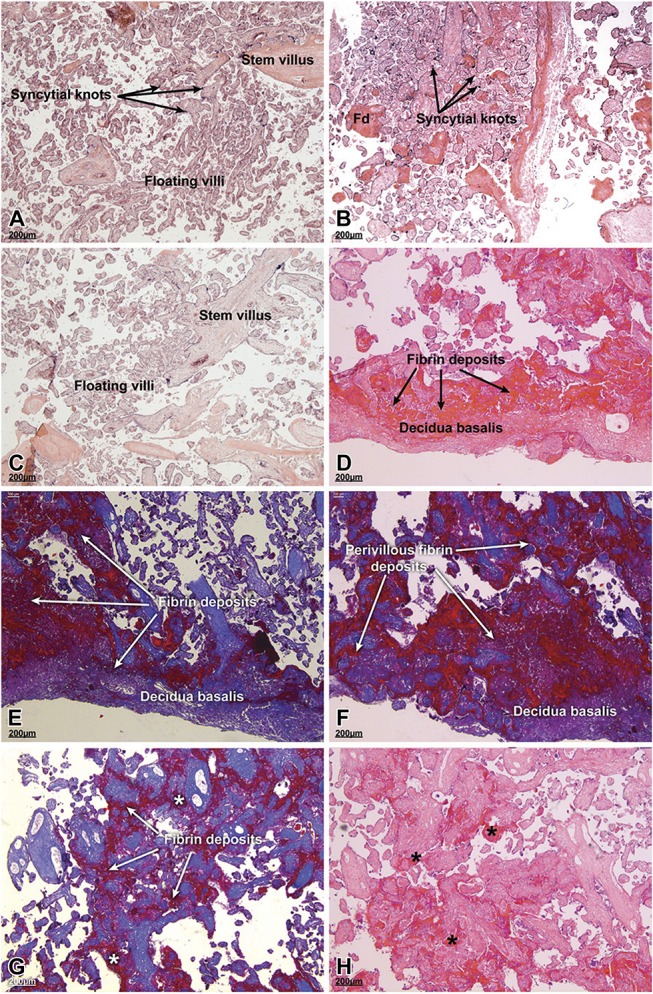
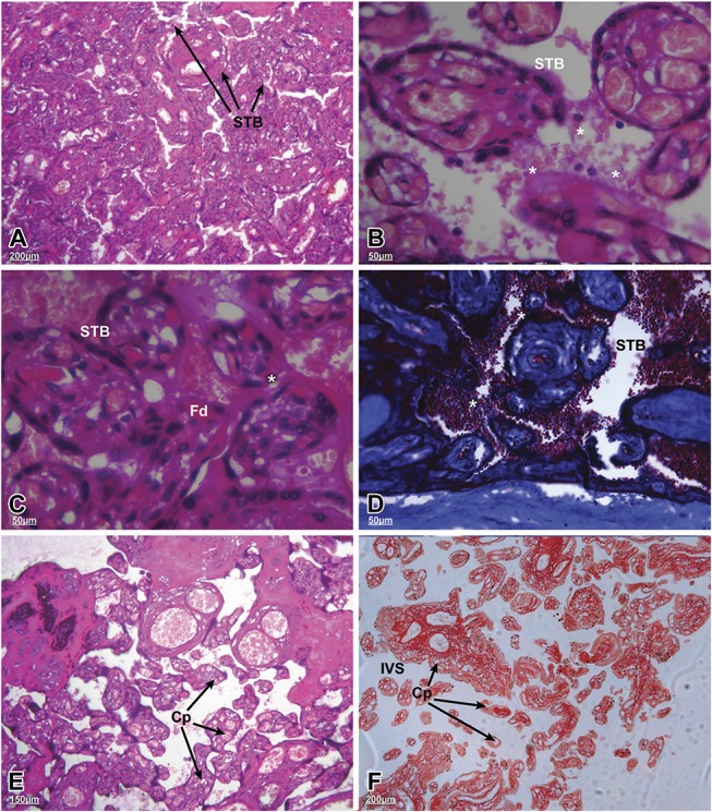
Results: Among preterm placentas, HIV infection was significantly associated with thrombosis (P = 0.001), infarction (P = 0.032), anomalies in cord insertion (P = 0.02), gross evidence of membrane infection (P = 0.043), and reduced placental thickness (P = 0.010). Overall, preterm placentas in both groups were associated with immature villi, syncytial knotting, villitis, and deciduitis. Features of HIV-positive versus HIV-negative placentas included significant fibrinoid deposition with villus degeneration, syncytiotrophoblast delamination, red blood cell adhesion, hypervascularity, and reduction in both surface area and perimeter of the terminal villi.

Conclusions: These results imply that HIV infection and/or ART are associated with morphological changes in preterm placentas that contribute to delivery before 37 weeks. Hypervascularity suggests that the observed pathologies may be attributable, in part, to hypoxia. Further research to explore potential mechanisms will help elucidate the pathways that are involved perhaps pointing to interventions for decreasing the risk of prematurity among HIV-positive women.


Conflict of interest statement

The authors have no conflicts of interest to disclose.

Figures

FIGURE 1.
FIGURE 1.
Histology of term and preterm placentas from HIV-positive women on antiretroviral treatment (ART) versus HIV-negative women. The placental biopsies were prepared for paraffin wax embedding, sectioned and stained with either hematoxylin and eosin (A–D and H) or Masson's trichrome (E–G) stains. A, Placenta from a normal HIV-negative pregnancy at term. Note the organization of the floating villi, stem villus, and the syncytial knots. Fibrin material is scant in this section. B, Placenta of a term pregnancy from a HIV-positive woman on ART. Numerous fibrin deposits (Fd) were evident as were syncytial knots, normal in a term placenta. C, Preterm placenta from a HIV-negative woman. The intervillous spaces, which were clear, contained intermediate mature and mature villi. D–H, Preterm placentas from HIV-positive women on ART had obvious fibrin deposition in the perivillous and intervillous regions sometimes obliterating the structure of floating and stem villi. Asterisks in panels G and H show areas of intervillous fibrin deposition. A, 39 weeks; (B) 39 weeks; (C) 33 weeks; (D) 34 weeks; (E) 35 weeks; (F) 34 weeks; (G) 33 weeks; and (H) 30 weeks of gestation. Scale bars, 200 μm.
FIGURE 2.
FIGURE 2.
Microstructural changes in the terminal villi of preterm placentas from HIV-positive women on antiretroviral therapy (ART). The sections were stained using hematoxylin and eosin (A–C, E and F) or Masson's trichrome (D). A, General architecture of the floating villi with their syncytiotrophoblast (STB) coverings (arrows; scale bar, 200 µm). B and C, Higher magnification of different fields from the same placenta showing regions of STB delamination associated with the terminal villi (asterisks). C, Areas of perivillous, eosinophilic fibrin deposits (Fd) were evident (scale bar 50 µm). D, Red blood cell adherence to the villi (asterisks; scale bar 50 µm). E and F, Higher number of capillaries (Cp) in the terminal villi with significant increases in the intervening intervillous spaces (IVS) [scale bars: (E) 150 µm; (F) 200 µm]. A, 35 weeks; (B) 35 weeks; (C) 35 weeks; (D) 32 weeks; (E) 33 weeks; and (F) 31 weeks of gestation.

References

    1. Lawn JE, Kinney M. Preterm birth: now the leading cause of child death worldwide. Sci Transl Med. 2014;6:263ed221. - PubMed
    1. Dekker GA, Lee SY, North RA, et al. Risk factors for preterm birth in an international prospective cohort of nulliparous women. PLoS One. 2012;7:e39154. - PMC - PubMed
    1. Butali A, Ezeaka C, Ekhaguere O, et al. Characteristics and risk factors of preterm births in a tertiary center in Lagos, Nigeria. Pan Afr Med J. 2016;24:1. - PMC - PubMed
    1. Rubens CE, Sadovsky Y, Muglia L, et al. Prevention of preterm birth: harnessing science to address the global epidemic. Sci Transl Med. 2014;6:262sr265. - PubMed
    1. Romero R, Dey SK, Fisher SJ. Preterm labor: one syndrome, many causes. Science. 2014;345:760–765. - PMC - PubMed

Publication types

MeSH terms