. 2019 Apr;13(4):84-91.
doi: 10.5489/cuaj.5142. Epub 2018 Sep 27.

18F-fluorocholine positron emission tomography-computed tomography (18F-FCH PET/CT) for staging of high-risk prostate cancer patients

Affiliations

18F-fluorocholine positron emission tomography-computed tomography (18F-FCH PET/CT) for staging of high-risk prostate cancer patients

Simon Gauvin et al. Can Urol Assoc J. 2019 Apr.

Abstract

Introduction: We sought to evaluate the diagnostic performance of 18F-fluorocholine positron emission tomography-computed tomography (18F-FCH PET/CT) for initial staging of patients with high-risk prostate cancer. Secondary objectives were to compare the value of 18F-FCH PET/CT to conventional imaging modalities and to evaluate its clinical impact.

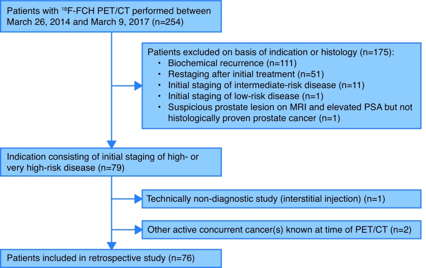
Methods: We conducted a retrospective study of 76 patients who underwent 18F-FCH PET/CT for initial staging of high-risk prostate cancer. Using pre-established validation criteria, sensitivity and specificity were determined for metastatic disease. Results were compared to findings on magnetic resonance imaging (MRI), computed tomography (CT), and bone scan (BS) when available.

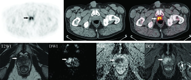
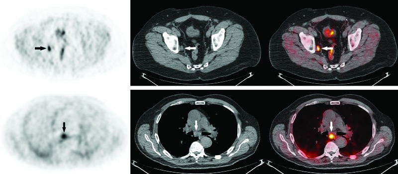
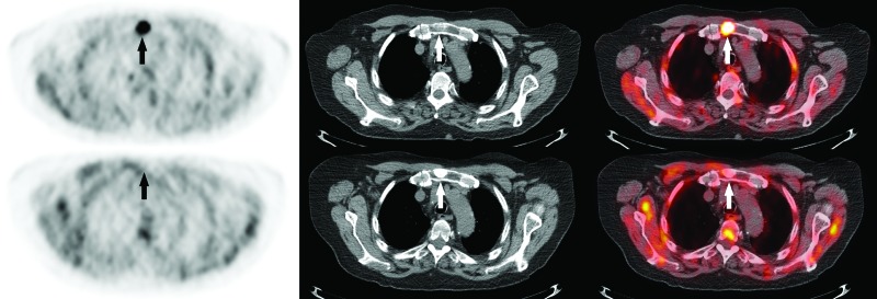
Results: Twenty-two (29%) PET/CT scans were positive, 49 (64%) negative, and five (7%) equivocal for nodal or metastatic disease. Of the positive scans, 17 showed regional lymph node involvement, 12 distant nodes, five bone metastases, and three lung metastases. Overall per-patient sensitivity, specificity, positive and negative predictive values for metastatic disease were 65%, 100%, 100%, and 78%, respectively. Sensitivity, specificity, and positive and negative predictive values were 64%, 100%, 100%, and 80%, respectively, for nodal involvement and 86%, 100%, 100%, and 98%, respectively, for bone and other metastases. Conventional imaging was negative for the lesion(s) found on PET/CT in five patients. PET/CT changed the clinical management in nine patients (12%).

Conclusions: Although 18F-FCH PET/CT offers some benefits over conventional imaging and demonstrates a high specificity, it remains limited by its sensitivity in the context of high-risk prostate cancer staging. PET with novel urea-based small molecule prostate-specific membrane antigen (PSMA) inhibitors may overcome some of these limitations. However, the interpretation of the study result is limited by the lack of available histological gold standard, the inclusion of several patients who received androgen-deprivation therapy (ADT) prior to PET/CT, our retrospective design, and a relatively small sample size.


Conflict of interest statement

Competing interests: This work was supported by the Departments of Nuclear Medicine and Urology, Jewish General Hospital, Montreal, QC, Canada. Dr. Probst has been an advisor for Bayer and has participated in a clinical trial supported by Progenics. The remaining authors report no competing personal or financial interests related to this work.

Figures

Fig. 1
Fig. 1
Flow diagram outlining inclusion and exclusion criteria and study design. 18F-FCH: 18F-fluorocholine; PET/CT: positron-emission tomography-computed tomography; MRI: magnetic resonance imaging; PSA: prostate-specific antigen.
Fig. 2
Fig. 2
Example of local disease on 18F-fluorocholine positron emission tomography-computed tomography (18F-FCH PET/CT) with magnetic resonance imaging (MRI) correlation. (A) Axial 18F-FCH PET/CT images obtained in a 63-year-old man with prostate cancer (cT1c, Gleason score 8, prostate-specific antigen 12.6 ng/mL) showing bilateral prostate uptake (SUV 6.6) (arrows) without metastatic disease. (B) Corresponding axial MRI prostate images (from left to right: T2-weighted images [T2WI], diffusion-weighted imaging [DWI], apparent diffusion coefficient [ADC] map, and dynamic contrast-enhanced images [DCE]) demonstrating a non-circumscribed homogeneous moderately T2 hypointense lesion measuring 1.8 cm in maximal dimension located in the transition zone at the apex and mid-gland with mild extension to the right anterolateral peripheral zone (arrows). There is associated restricted diffusion on DWI/ADC and early focal enhancement on DCE (PI-RADS 5). The patient underwent radical prostatectomy with extended pelvic lymph node dissection (pT3a pN0), without evidence of biochemical recurrence after 10 months of followup. MRI images courtesy of Dr. F. Discepola, Jewish General Hospital, Montreal, QC, Canada.
Fig. 3
Fig. 3
Examples of regional and distant lymph node involvement on 18F-fluorocholine positron emission tomography-computed tomography (18F-FCH PET/CT). (A) Axial 18F-FCH PET/CT images obtained in a 68-year-old man with prostate cancer (PCa) (cT1a, Gleason score 9, prostate-specific antigen [PSA] 17.2 ng/mL) showing intense 18F-FCH uptake (SUV 6.7) in a 1.1 cm right obturator lymph node (arrows). The patient underwent radical prostatectomy with pelvic lymph node dissection (pT3a pN1). (B) Axial 18F-FCH PET/CT images obtained in a 78-year-old man with metastatic PCa (cT3b, Gleason score 9, PSA 11.1 ng/mL) showing abnormal uptake in a subcarinal lymph node (arrows), proven to represent PCa metastasis on endobronchial ultrasound-guided transbronchial needle aspiration biopsy (EBUS-TBNA).
Fig. 4
Fig. 4
Example of bone metastasis on 18F-fluorocholine positron emission tomography-computed tomography (18F-FCH PET/CT). (A) Axial 18F-FCH PET/CT images obtained at initial staging in a 82-year-old man with prostate cancer (cT3b, Gleason score 6, prostate-specific antigen 81.0 ng/mL) showing focal intense manubrial 18F-FCH uptake (SUV 11.8) (arrows). (B) Followup PET/CT obtained four months after androgen-deprivation therapy demonstrates complete metabolic response of the manubrial lesion, which is now densely sclerotic (arrows).

References

    1. Cancer.ca. Canadian Cancer Society’s Advisory Committee on Cancer Statistics. 2017. [Accessed May 19, 2018]. [updated 2017; cited 2018 May 19]. Available at: http://www.cancer.ca/
    1. D’Amico AV, Whittington R, Malkowicz SB, et al. Biochemical outcome after radical prostatectomy, external beam radiation therapy, or interstitial radiation therapy for clinically localized prostate cancer. JAMA. 1998;280:969–74. doi: 10.1001/jama.280.11.969. - DOI - PubMed
    1. Cooperberg MR, Cowan J, Broering JM, et al. High-risk prostate cancer in the United States, 1990–2007. World J Urol. 2008;26:211–8. doi: 10.1007/s00345-008-0250-7. - DOI - PMC - PubMed
    1. Weiner AB, Matulewicz RS, Eggener SE, et al. Increasing incidence of metastatic prostate cancer in the United States (2004–2013) Prostate Cancer Prostatic Dis. 2016;19:395–97. doi: 10.1038/pcan.2016.30. - DOI - PubMed
    1. Sanda MG, Chen RC, Crispino T, et al. 2017. [Accessed Nov 24, 2017]. [cited 2017 November 24]. Available at: http://www.auanet.org/guidelines/clinically-localized-prostate-cancer-ne...