. 2018 Sep 28;19(10):2957.
doi: 10.3390/ijms19102957.

Inhibitory Antibodies against Activin A and TGF-β Reduce Self-Supported, but Not Soluble Factors-Induced Growth of Human Pulmonary Arterial Vascular Smooth Muscle Cells in Pulmonary Arterial Hypertension

Affiliations

Inhibitory Antibodies against Activin A and TGF-β Reduce Self-Supported, but Not Soluble Factors-Induced Growth of Human Pulmonary Arterial Vascular Smooth Muscle Cells in Pulmonary Arterial Hypertension

Tatiana V Kudryashova et al. Int J Mol Sci. .

Abstract

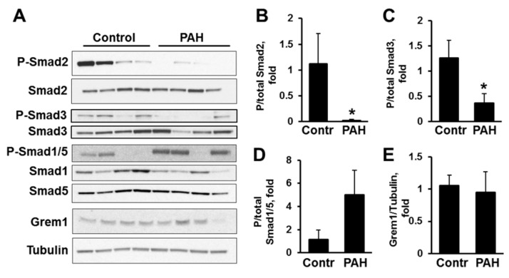
Increased growth and proliferation of distal pulmonary artery vascular smooth muscle cells (PAVSMC) is an important pathological component of pulmonary arterial hypertension (PAH). Transforming Growth Factor-β (TGF-β) superfamily plays a critical role in PAH, but relative impacts of self-secreted Activin A, Gremlin1, and TGF-β on PAH PAVSMC growth and proliferation are not studied. Here we report that hyper-proliferative human PAH PAVSMC have elevated secretion of TGF-β1 and, to a lesser extent, Activin A, but not Gremlin 1, and significantly reduced Ser465/467-Smad2 and Ser423/425-Smad3 phosphorylation compared to controls. Media, conditioned by PAH PAVSMC, markedly increased Ser465/467-Smad2, Ser423/425-Smad3, and Ser463/465-Smad1/5 phosphorylation, up-regulated Akt, ERK1/2, and p38 MAPK, and induced significant proliferation of non-diseased PAVSMC. Inhibitory anti-Activin A antibody reduced PAH PAVSMC growth without affecting canonical (Smads) or non-canonical (Akt, ERK1/2, p38 MAPK) effectors. Inhibitory anti-TGF-β antibody significantly reduced P-Smad3, P-ERK1/2 and proliferation of PAH PAVSMC, while anti-Gremlin 1 had no anti-proliferative effect. PDGF-BB diminished inhibitory effects of anti-Activin A and anti-TGF-β antibodies. None of the antibodies affected growth and proliferation of non-diseased PAVSMC induced by PAH PAVSMC-secreted factors. Together, these data demonstrate that human PAH PAVSMC have secretory, proliferative phenotype that could be targeted by anti-Activin A and anti-TGF-β antibodies; potential cross-talk with PDGF-BB should be considered while developing therapeutic interventions.

Keywords: Activin A; Gremlin 1; PDGF-BB; Smad proteins; TGF-β; growth; human smooth muscle cells; proliferation; pulmonary arterial hypertension; therapeutic antibody.


Conflict of interest statement

The authors declare no conflict of interest.

Figures

Figure 1
Figure 1
Human pulmonary arterial hypertension (PAH) pulmonary arterial vascular smooth muscle cells (PAVSMC) have increased secretion of transforming growth factor-β (TGF-β1). Human non-diseased (Control) and PAH PAVSMC were incubated for 48 h in cultural media supplemented with 0.1% bovine serum albumin (BSA); then media was collected and protein levels of TGF-β1 (A), Activin A (B), and Gremlin 1 (Grem1) (C) were measured in conditioned media by quantitative sandwich enzyme-linked immunosorbent assay (ELISA) (A,B) or immunoblot analysis (C). Data are means ± SE; * p < 0.05 by Mann-Whitney U test vs. control; n = 3–4 subjects/group. N-S—non-specific.
Figure 2
Figure 2
Human PAH PAVSMC have reduced phosphorylation of Smad2 and Smad3. (A) Human non-diseased (Control) and PAH PAVSMC were incubated for 48 h in cultural media with 0.1% BSA followed by immunoblot analysis to detect indicated proteins; (BE) Data are means ± SE; * p < 0.05 by Mann-Whitney U test vs. control; n = 4 subjects/group.
Figure 3
Figure 3
Effect of inhibitory antibodies to Activin A, Gremlin 1 and TGF-β on growth of human non-diseased and PAH PAVSMC. (AC) Cells were maintained in changed daily serum-free media supplemented with 3.5 nM of indicated antibodies in the presence or absence (diluent) of 10 ng/mL PDGF-BB (A,B) or 1 µM bosentan or diluent (C); cell counts were performed at days 0, 3, and 5. Data are means ± SE representing fold to day 0 from 3 subjects/groups. (D) Cells, serum-deprived for 48 h, were treated with 3.5 nM of indicated antibodies in the presence or absence (diluent) of 10 ng/mL PDGF-BB for 24 h and then DNA synthesis was examined using BrdU incorporation assay. Data are means ± SE fold to control; * p < 0.05 by Mann-Whitney U test; n = 3 subjects/group.
Figure 4
Figure 4
Effect of inhibitory antibodies to Activin A, Gremlin 1 and TGF-β on Smad phosphorylation status in human non-diseased and PAH PAVSMC. Cells, serum-deprived for 48 h, were treated with 3.5 nM antibodies to Activin A, Gremlin 1 and TGF-β or control IgG in the presence or absence of 10 ng/mL PDGF-BB for 18 h, and immunoblot analysis to detect indicated proteins was performed. (A) Representative immunoblots from three experiments, each performed on the cells from different human subject; (B) Data represent fold changes in P/total protein ratios with P/total ratio for control IgG without stimulation taken as 1 fold. Data are means ± SE; * p < 0.05 by Mann-Whitney U test; n = 3 subjects/group.
Figure 5
Figure 5
Effect of inhibitory antibodies to Activin A, Gremlin 1 and TGF-β on pro-proliferative signaling molecules in human PAH PAVSMC. Cells, serum-deprived for 48 h, were treated with 3.5 nM antibodies to Activin A, Gremlin 1 and TGF- β, or control IgG in the presence or absence of 10 ng/mL PDGF-BB for 18 h, and immunoblot analyses to detect phosphorylation status of Akt (A), extracellular signal-regulated kinases ½ (ERK1/2) (B), and p38 mitogen-activated protein kinase (MAPK) (C) were performed. Top panels: Representative immunoblots from three experiments, each performed on the cells from different human subject. Bottom panels: Data represent fold changes in P/total protein ratios with P/total ratio for control IgG without stimulation taken as 1 fold. Data are means ± SE, * p < 0.05 by Mann-Whitney U test; n = 3 subjects/group.
Figure 6
Figure 6
PAH PAVSMC conditioned medium promotes proliferation of non-diseased human PAVSMC. (A) Experimental design: Conditioned medium from serum-deprived non-diseased (Contr CM) and PAH PAVSMCs (PAH CM) was harvested after 48 h of incubation. Non-diseased cells were serum-deprived for 48 h, incubated with Contr CM or PAH CM for 24 h followed by DNA synthesis analysis using BrdU incorporation assay. Non-stimulated cells and cells treated with 10 ng/mL PDGF-BB were used as a negative and positive control, respectively. (B) Data are means ± SE; Data are folds to negative control; * p < 0.05 by Mann-Whitney U test vs. control; n = 4 subjects/group.
Figure 7
Figure 7
Media, conditioned by PAH PAVSMC, promotes Smad, Akt, ERK1/2 and p38 MAPK phosphorylation in non-diseased human PAVSMC. Conditioned medium from serum-deprived non-diseased (Contr CM) and PAH PAVSMCs (PAH CM) was harvested after 48 h of incubation. Non-diseased cells were serum-deprived for 48 h, incubated with Contr CM or PAH CM for 24 h, and immunoblot analysis to detect indicated proteins was performed. Non-stimulated cells and cells treated with 10 ng/mL PDGF-BB were used as a negative and positive control, respectively. (A) Representative immunoblots from three experiments, each performed on the cells from different human subject; (B,C) Data are means ± SE; Data are P/total ratios represented as a folds to negative control; * p < 0.05 by Mann-Whitney U test vs. control; n = 3 subjects/group.
Figure 8
Figure 8
Inhibitory antibodies against Activin A, Gremlin 1 and TGF-β have no significant effect on the growth of human non-diseased PAVSMC induced by PAH PAVSMC-conditioned media. (A) Non-diseased (control) human PAVSMC were maintained in changed daily serum-free media supplemented with 3.5 nM of indicated antibodies in the presence of conditioned media collected after 48 h of incubation with serum-deprived control (Contr CM) or PAH PAVSMCs (PAH CM), and cell counts were performed at days 0, 3, and 5. Data are means ± SE presented as a folds to day 0; n = 3 subjects/group. * p < 0.05 by Mann-Whitney U test vs control. (B) Control human PAVSMC were serum deprived for 48 h, treated with 3.5 nM of indicated antibodies in the presence of Contr CM or PAH CM for 24 h, incubated with BrdU for 18 h, and DNA synthesis was examined using BrdU incorporation assay. Data are means ± SE presented as fold to control from 5 subjects/group. * p < 0.05 by Mann-Whitney U test vs control. (C) Control human PAVSMC were serum-deprived, treated with 10 ng/mL TGF-β1, 10 ng/mL PDGF-BB, or diluent for 5 days, and DNA synthesis analysis was performed using BrdU incorporation assay. Cells from two different subjects were analyzed.

References

    1. Humbert M., Sitbon O., Chaouat A., Bertocchi M., Habib G., Gressin V., Yaici A., Weitzenblum E., Cordier J.F., Chabot F., et al. Survival in patients with idiopathic, familial, and anorexigen-associated pulmonary arterial hypertension in the modern management era. Circulation. 2010;122:156–163. doi: 10.1161/CIRCULATIONAHA.109.911818. - DOI - PubMed
    1. Benza R.L., Miller D.P., Barst R.J., Badesch D.B., Frost A.E., McGoon M.D. An evaluation of long-term survival from time of diagnosis in pulmonary arterial hypertension from the reveal registry. Chest. 2012;142:448–456. doi: 10.1378/chest.11-1460. - DOI - PubMed
    1. Galie N., Manes A., Negro L., Palazzini M., Bacchi-Reggiani M.L., Branzi A. A meta-analysis of randomized controlled trials in pulmonary arterial hypertension. Eur. Heart J. 2009;30:394–403. doi: 10.1093/eurheartj/ehp022. - DOI - PMC - PubMed
    1. Frost A.E., Badesch D.B., Barst R.J., Benza R.L., Elliott C.G., Farber H.W., Krichman A., Liou T.G., Raskob G.E., Wason P. The changing picture of patients with pulmonary arterial hypertension in the united states: How reveal differs from historic and non-us contemporary registries. Chest. 2011;139:128–137. doi: 10.1378/chest.10-0075. - DOI - PubMed
    1. Humbert M., Morrell N.W., Archer S.L., Stenmark K.R., MacLean M.R., Lang I.M., Christman B.W., Weir E.K., Eickelberg O., Voelkel N.F., et al. Cellular and molecular pathobiology of pulmonary arterial hypertension. J. Am. Coll. Cardiol. 2004;43:S13–S24. doi: 10.1016/j.jacc.2004.02.029. - DOI - PubMed

MeSH terms