. 2018 Sep 28;10(10):363.
doi: 10.3390/cancers10100363.

Fenofibrate Interferes with the Diapedesis of Lung Adenocarcinoma Cells through the Interference with Cx43/EGF-Dependent Intercellular Signaling

Affiliations

Fenofibrate Interferes with the Diapedesis of Lung Adenocarcinoma Cells through the Interference with Cx43/EGF-Dependent Intercellular Signaling

Katarzyna Piwowarczyk et al. Cancers (Basel). .

Abstract

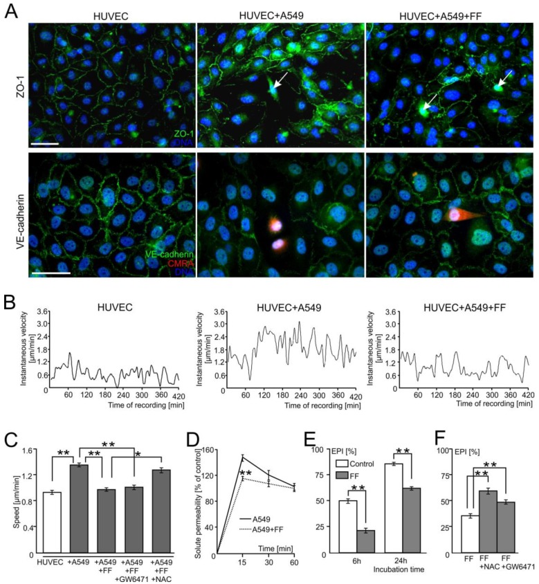
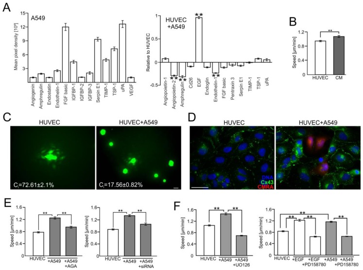
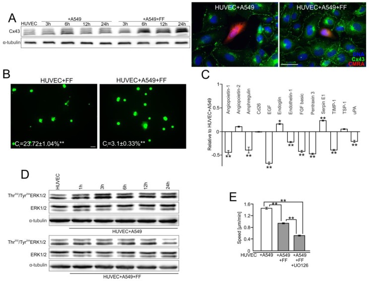
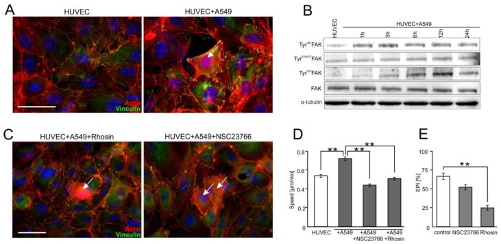
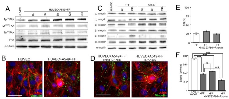
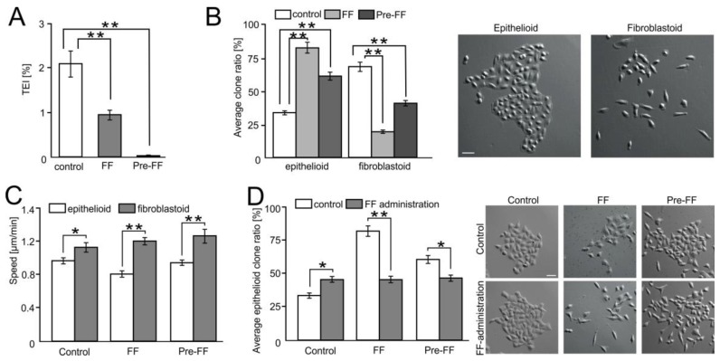
Extravasation of circulating cancer cells is regulated by the intercellular/intracellular signaling pathways that locally impair the endothelial barrier function. Co-cultures of human umbilical vein endothelial cells (HUVECs) with lung adenocarcinoma A549 cells enabled us to identify these pathways and to quantify the effect of fenofibrate (FF) on their activity. A549 cells induced the disruption and local activation of endothelial continuum. These events were accompanied by epidermal growth factor (EGF) up-regulation in endothelial cells. Impaired A549 diapedesis and HUVEC activation were seen upon the chemical inhibition of connexin(Cx)43 functions, EGF/ERK1/2-dependent signaling, and RhoA/Rac1 activity. A total of 25 μM FF exerted corresponding effects on Cx43-mediated gap junctional coupling, EGF production, and ERK1/2 activation in HUVEC/A549 co-cultures. It also directly augmented endothelial barrier function via the interference with focal adhesion kinase (FAK)/RhoA/Rac1-regulated endothelial cell adhesion/contractility/motility and prompted the selective transmigration of epithelioid A549 cells. N-acetyl-L-cysteine abrogated FF effects on HUVEC activation, suggesting the involvement of PPARα-independent mechanism(s) in its action. Our data identify a novel Cx43/EGF/ERK1/2/FAK/RhoA/Rac1-dependent signaling axis, which determines the efficiency of lung cancer cell diapedesis. FF interferes with its activity and reduces the susceptibility of endothelial cells to A549 stimuli. These findings provide the rationale for the implementation of FF in the therapy of malignant lung cancers.

Keywords: Cx43; EGF; diapedesis; fenofibrate; lung cancer.


Conflict of interest statement

The authors declare no conflict of interest.

Figures

Figure 1
Figure 1
Fenofibrate inhibits the penetration of the endothelial layer by lung adenocarcinoma (A549) cells. Human lung cancer A549 cells (stained with CMRA-Cell Tracker or marked with arrows) were seeded onto human umbilical vein endothelial cell (HUVEC) monolayers at the density of 1300 cells/cm2. The integrity of endothelial continua, HUVEC motility, and endothelial permeability was assessed by anti-ZO-1/VE-cadherin immunostaining ((A); 6 h), time-lapse videomicroscopy ((B,C); 7 h), and solute permeability tests (D), respectively, in the absence or in the presence of 25 μM fenofibrate (FF). (B,C) shows instantaneous speeds of single HUVEC in control conditions and in the presence of A549/25 μM FF, and the comparison of average speeds of HUVEC movement in the presence of A549 cells/FF/GW6471/NAC, respectively, quantified based on changes in cell centroid position. (E) The effect of FF on the efficiency of A549 diapedesis (TransEndothelial Penetration; EPI) was estimated after 6 and 24 h of co-culture. (F) Effect of PPARα inhibitor (GW6471) and ROS scavenger (N-acetyl-L-cysteine; NAC) on A549 diapedesis (EPI), estimated after 6 h of A549/HUVEC co-culture. Transmigration of at least 200 A549 cells was analyzed for each group. The statistical significance of the differences in (CF) was tested with one-way ANOVA followed by a post-hoc Tukey’s HSD comparison (* p < 0.05 and ** p < 0.01). Error bars represent SEM. All results are representative of at least three independent experiments (n ≥ 3). Scale bar = 40 μm. Note that the relatively efficient diapedesis of A549 cells is considerably inhibited by FF.
Figure 2
Figure 2
A549 cells impair the endothelial barrier function via the activation of the Cx43/EGF/ERK1/2-dependent intercellular signaling axis. (A) A549 cells were seeded onto HUVEC monolayers as in Figure 1 and co-cultured for 24 h. Then, the expression of angioactive proteins was semi-quantitively estimated with an antibody array kit (see Materials and Methods). Plots show the densitometrically estimated dot intensities, illustrating the protein amounts in A549 cells (in a.u.; left) or in A549/HUVEC co-cultures relative to the HUVEC control. (B) A549-conditioned medium (3:5) was added to HUVECs and their motility was estimated with time-lapse videomicroscopy for 7 h. (C) Calcein-loaded HUVEC (left) or A549 cells (right) were seeded onto HUVEC monolayers and GJIC (coupling ratio-Ci) was estimated by a calcein transfer assay after 1 h. Concomitantly, Cx43 expression in HUVECs and in HUVEC/A549 co-cultures was estimated with immunofluorescence (D). (E) The effect of AGA (70 μM) and Cx43 silencing by siRNA on HUVEC motility. (F) HUVECs were cultured in the presence of EGF or A549/HUVEC co-cultures were established as above and the effects of EGFR- or ERK1/2 inhibitor (PD158780 and UO126, respectively) on HUVEC motility were estimated with time-lapse videomicroscopy. Error bars represent SEM. Scale bar = 40 μm. The statistical significance of the differences was tested with one-way ANOVA followed by post-hoc Tukey’s HSD (B,E) or non-parametric Dunnett comparison (A,F); ** p < 0.01. All results are representative of at least three independent experiments (n ≥ 3). Note the presence of EGF in A549/HUVEC co-cultures and the attenuating effect of chemical Cx43/EGFR and ERK1/2 inhibition on A549-induced HUVEC activation.
Figure 3
Figure 3
Fenofibrate interferes with the communication networks established between HUVEC and A549 cells. (A) HUVEC/A549 co-cultures were established as in Figure 1 and cultivated in the absence or presence of 25 μM FF. Cx43 levels were analyzed at the indicated time points by immunoblotting, quantified by densitometry (Figure S5A,B) and visualized by immunofluorescence after 6 h. (B) Calcein-loaded HUVEC (left) or A549 cells (right) were seeded onto HUVEC monolayers and the effect of FF on GJIC (coupling ratio-Ci) was estimated by calcein transfer assay after 1 h. (C) Expression of angioactive proteins in FF-treated HUVEC/A549 co-culture was semi-quantitively estimated by the antibody array kit. Plots show the densitometrically estimated dot intensities, illustrating the amounts of a given factor in HUVEC/A549 cells in the presence of 25 μM FF (24 h; relative to HUVEC/A549 control). (D) HUVEC/A549 co-cultures were cultivated in the absence or in the presence of 25 μM FF. Tyr202/204ERK1/2 levels were analyzed at the indicated time points by immunoblotting and quantified by densitometry (Figure S5C). (E) HUVEC/A549 co-cultures were established as above and cultivated in the presence of 25 μM FF. The effect of the ERK1/2 inhibitor (UO126) on HUVEC activation was analyzed with time-lapse videomicroscopy. Error bars represent SEM. Scale bar = 40 μm. The statistical significance of the differences was tested with one-way ANOVA followed by post-hoc Tukey’s HSD (B) or non-parametric Dunnett comparison (AE); * p < 0.05; ** p < 0.01. All results are representative of a least three independent experiments (n ≥ 3). Note that FF administration inhibits GJIC between cancer and endothelial cells, abolishes the induction of EGF secretion, and inhibits A549-induced motility of HUVECs.
Figure 4
Figure 4
Focal adhesion kinase (FAK)/RhoA-dependent signaling participates in the activation of HUVECs by A549 cells. (A) A549 cells (marked with arrow) were seeded onto the monolayer of HUVEC as described in Figure 1, cultivated for 6 h, fixed, and stained for F-actin and vinculin. (B) A549/HUVEC co-cultures were established as above, and Tyr397, Tyr576/577, and Tyr925 FAK were analyzed at the indicated time points by immunoblotting and quantified by densitometry (Figure S5D). (C) Cytoskeletal architecture of HUVEC cultured in co-cultures with A549 cells treated with Rhosin (left) or NSC23766 for 6 h (right). (D,E) A549/HUVEC co-cultures were treated with Rhosin and NSC23766 followed by the analyses of HUVEC motility (D) and A549 diapedesis ((E); 6 h). Error bars represent SEM. The statistical significance of the differences was tested with one-way ANOVA followed by post-hoc Tukey’s HSD (D,E); ** p < 0.01. All results are representative of at least three independent experiments (n ≥ 3). Scale bar = 40 μm. Note that RhoA/Rac1-dependent disruption of endothelial continuum by A549 cells is accompanied by FAK activation and cytoskeletal rearrangements in proximal HUVECs.
Figure 5
Figure 5
FF directly attenuates HUVEC susceptibility to the activating signals generated by A549 cells. (A) HUVEC/A549 co-cultures were established as in Figure 1 and cultivated in the presence of 25 μM FF. Tyr397FAK, Tyr576/577FAK, and Tyr925FAK were analyzed at the indicated time points by immunoblotting and quantified by densitometry (Figure S5E). (B) Cytoskeletal architecture in HUVECs (Figure 3A) in A549/HUVEC co-cultures visualized by immunostaining in the absence (left) or presence of 25 μM FF (6 h; right). (C) HUVECs were cultivated in the presence of 25 μM FF or A549 cells and the levels of integrins were estimated at the indicated time points by immunoblotting and quantified by densitometry (Figure S5F). (D) The effect of NSC23766 (left) or Rhosin (right) on the cytoskeletal architecture of HUVECs cultured in the presence of A549 cells and 25 μM FF (see B for control). (E) Effect of NSC23766, Rhosin, and FF on the efficiency of A549 diapedesis (TransEndothelial Penetration; EPI), estimated after 6 h of A549/HUVEC co-culture. Transmigration of at least 200 A549 cells was analyzed for each group. (F) Effect of NSC23766 and Rhosin on the motility of proximal HUVECs analyzed in the presence of FF by time-lapse videomicroscopy. Error bars represent SEM. The statistical significance of the differences was tested with one-way ANOVA followed by post-hoc Tukey’s HSD (E) or non-parametric Dunnett comparison (F); * p < 0.05; ** p < 0.01 against the control or # p < 0.05 against HUVEC/A549 control (see Figure 4B and Figure S5D). All results are representative of at least three independent experiments (n ≥ 3). Scale bar = 40 μm. Note that the inhibition of RhoA and Rac1 slightly counteracts FF-induced maturation of focal adhesions while inhibiting A549 diapedesis and HUVEC motility.
Figure 6
Figure 6
FF selectively impairs the transendothelial migration of fibroblastoid A549 cells. (A) A549 cells were seeded on microporous membranes covered by control or FF-pretreated (Pre-FF) HUVEC monolayers, cultivated for 24 h in control conditions or in the presence of 25 μM FF (FF), and the fraction of transmigrated cells was estimated (TransEndothelial penetration Index (TEI)). (B) Morphology of A549 microclones derived from the cells that transmigrated through HUVEC-covered microporous membranes in the same conditions as in (A). (C) Motility of A549 progenies derived from the cells that transmigrated through HUVEC-covered microporous membranes in the same conditions as in (A). (D) Progenies derived from A549 cells that most readily transmigrated through HUVEC monolayers were treated with FF, and their morphology and the fraction of epithelioid clones was estimated after 72 h. Error bars represent SEM. The statistical significance of the differences was tested with one-way ANOVA followed by post-hoc Tukey’s HSD (A,B,D) or non-parametric Dunnett comparison (C); * p < 0.05; ** p < 0.01. All results are representative of at least three independent experiments (n ≥ 3). Scale bar = 40 μm. Note the differential effect of FF on the transmigration of “fibroblastoid” and “epithelioid” A549 cells.

References

    1. Siegel R.L., Miller K.D., Jemal A. Cancer statistics, 2016. CA Cancer J. Clin. 2016;66:7–30. doi: 10.3322/caac.21332. - DOI - PubMed
    1. Torre L.A., Siegel R.L., Jemal A. Lung Cancer Statistics. Adv. Exp. Med. Biol. 2016;893:1–19. doi: 10.1007/978-3-319-24223-1_1. - DOI - PubMed
    1. Savagner P. The epithelial-mesenchymal transition (EMT) phenomenon. Ann. Oncol. 2010;21(Suppl. 7):vii89–vii92. doi: 10.1093/annonc/mdq292. - DOI - PMC - PubMed
    1. Wang X., Ferreira A.M., Shao Q., Laird D.W., Sandig M. Beta3 integrins facilitate matrix interactions during transendothelial migration of PC3 prostate tumor cells. Prostate. 2005;63:65–80. doi: 10.1002/pros.20168. - DOI - PubMed
    1. Woodward J. Crossing the endothelium: E-selectin regulates tumor cell migration under flow conditions. Cell Adhes. Migr. 2008;2:151–152. doi: 10.4161/cam.2.3.6820. - DOI - PMC - PubMed